Highlight
งานวิจัยนี้รายงานความแปรผันทางกายวิภาคของช่องกระดูกขากรรไกรแบบไม่สมบูรณ์ทั้งสองข้างที่ไม่เคยมีรายงานมาก่อน โดยศึกษาโครงสร้างทางกายวิภาคและการพัฒนาของช่องดังกล่าวผ่านภาพเอกซเรย์คอมพิวเตอร์และแบบจำลองสามมิติ ซึ่งมีผลต่อการตีความภาพรังสีและการวางแผนผ่าตัดขากรรไกรล่าง
ที่มาและความสำคัญ
ช่องกระดูกขากรรไกร (mandibular canal) เป็นช่องที่สำคัญในการส่งผ่านเส้นประสาทและหลอดเลือดในขากรรไกรล่าง แต่รูปแบบความแปรผันของช่องกระดูกขากรรไกรยังมีรายงานไม่มากนัก การศึกษานี้รายงานความแปรผันทางกายวิภาคที่ไม่เคยอธิบายมาก่อนของช่องกระดูกขากรรไกรแบบไม่สมบูรณ์ทั้งสองข้างในกระดูกขากรรไกรจากซากศพชาย ด้วยการตรวจทางกายวิภาคและภาพเอกซเรย์คอมพิวเตอร์ จากนั้นใช้ชิ้นเนื้อจากตัวอ่อนมนุษย์เพื่อสร้างแบบจำลองสามมิติ วิเคราะห์ความสัมพันธ์ระหว่างกระดูกขากรรไกรที่กำลังพัฒนา เส้นประสาทขากรรไกรล่าง และต่อมน้ำลาย ผลการศึกษาสนับสนุนสมมติฐานว่า การก่อตัวของช่องที่ไม่สมบูรณ์อาจเกิดจากตำแหน่งของเส้นประสาทหรือการถูกกักขังของเนื้อเยื่อต่อมน้ำลายในระหว่างการพัฒนา ความแปรผันนี้มีความสำคัญต่อการตีความภาพทางรังสีและการวางแผนผ่าตัดขากรรไกรล่างในทางคลินิก
Abstract
Purpose
This study aims to report a previously undocumented anatomical variation of the mandible and investigate its developmental origin using embryological analysis.
Methods
Anatomical inspection and computed tomography were performed on a dried human mandible from a male cadaver of unknown age. Additionally, serial histological sections from a Carnegie Stage 23 human embryo were reconstructed in 3D model to examine the spatial relationships between the developing mandible and surrounding structures.
Results
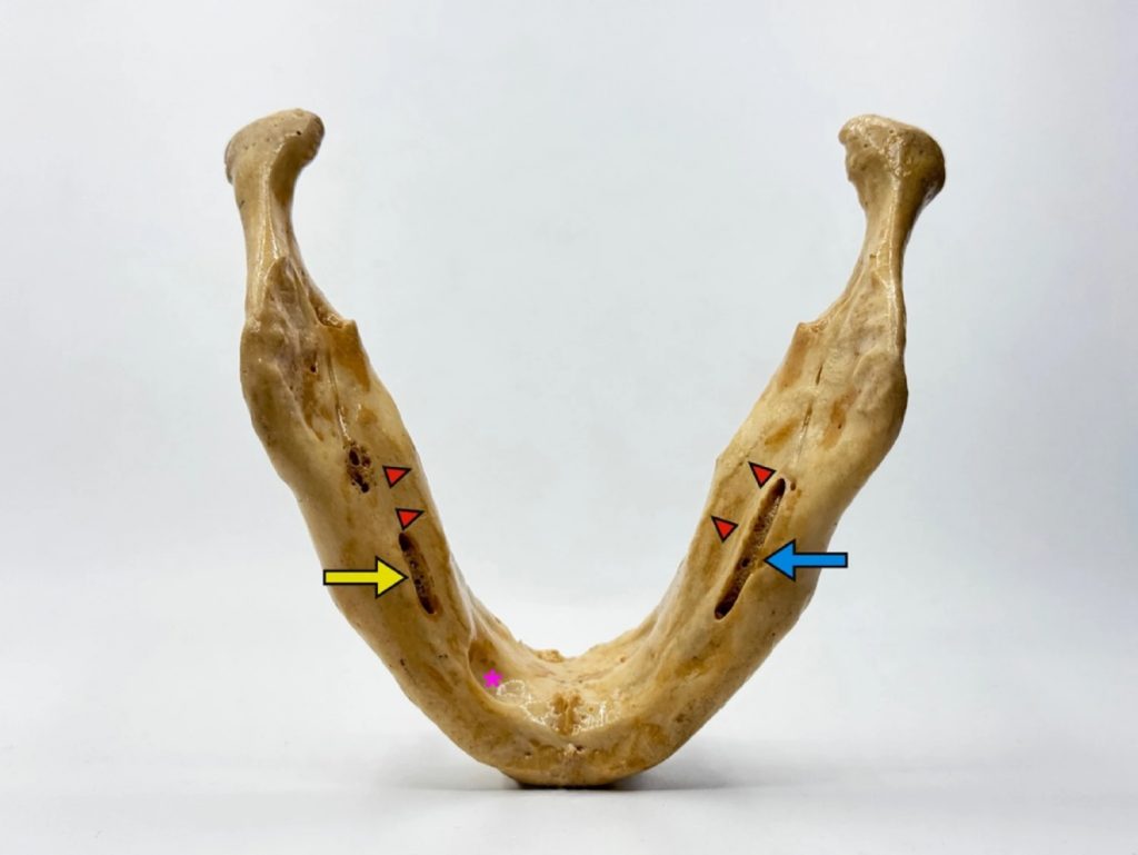
The mandibular canal exhibited breaching of the medial wall into the lingual cortex on both sides. Computed tomography confirmed that the canals originated from the mandibular foramen and gave rise to the mental foramina at the medial third of the mandible. These defects were found along with a left-sided Stafne bone cavity. Embryological analysis revealed a close spatial proximity between the developing mandible, the inferior alveolar nerve, and the salivary glands, supporting the hypothesis that incomplete ossification could result from variant nerve positioning or glandular entrapment.
Conclusion
This study presents rare anatomical variations of the mandibular canal and their possible developmental mechanism. These findings have implications for radiological interpretation and surgical planning involving the mandible.
KEYWORDS: Mandible, Mandibular canal, Anatomical variation, Computed tomography, Embryological analysis
Citation: Rumpansuwon, K., Berkban, T., Kruepunga, N. et al. Bilateral incomplete mandibular canals: an embryological analysis of their possible etiology. Surg Radiol Anat47, 233 (2025). https://doi.org/10.1007/s00276-025-03749-y
RELATED SDGs:
3. GOOD HEALTH AND WELL-BEING

ผู้ให้ข้อมูล: ผู้ช่วยศาสตราจารย์ ดร.อธิคุณ สุวรรณขันธ์
ชื่ออาจารย์ที่ทำวิจัย: รองศาสตราจารย์ ดร.วัฒนา วีรชาติยานุกูล รองศาสตราจารย์ ดร.สมลักษณ์ อสุวพงษ์พัฒนา ผู้ช่วยศาสตราจารย์ ดร.อธิคุณ สุวรรณขันธ์
ชื่อนักศึกษาที่ทำวิจัย: นายคณิติน รำแพนสุวรรณ
Credit ภาพ: ผู้ช่วยศาสตราจารย์ ดร.อธิคุณ สุวรรณขันธ์
Webmaster: ว่าที่ ร.อ. นเรศ จันทรังสิกุล
Tags: anatomical variation, computed tomography, Embryological analysis, Mandible, Mandibular canal