Highlight
การศึกษาพบว่าความแปรผันของฐานกะโหลกศีรษะส่วน clivus พบได้บ่อย รวมถึงร่อง fossa navicularis magna, ช่อง canalis basilaris medianus, และตุ่มที่ขอบ foramen magnum ข้อมูลเหล่านี้ช่วยเพิ่มความแม่นยำในการตีความภาพเอกซเรย์คอมพิวเตอร์และการวางแผนทางศัลยกรรมฐานกะโหลกศีรษะในผู้ป่วยชาวไทย
ที่มาและความสำคัญ
การแปรผันทางกายวิภาคของฐานกะโหลกศีรษะส่วน clivus เป็นข้อมูลสำคัญสำหรับการวินิจฉัยทางรังสีและการผ่าตัดศีรษะ–คอ เนื่องจากตำแหน่งที่อยู่ใกล้กับโครงสร้างประสาทและหลอดเลือดสำคัญ แต่มีข้อมูลเกี่ยวกับความแปรผันเหล่านี้ในประชากรไทยค่อนข้างจำกัด งานวิจัยนี้ใช้ภาพถ่ายเอกซเรย์คอมพิวเตอร์จากคนไทยมากกว่า 400 รายเพื่อประเมินความชุกและลักษณะของความแปรผันหลายแบบ เช่น ร่อง fossa navicularis magna ช่อง canalis basilaris medianus และตุ่มที่ขอบ foramen magnum ผลการศึกษาพบว่าความแปรผันเหล่านี้พบได้ค่อนข้างบ่อยที่สุดคือร่อง fossa navicularis magna ซึ่งมีความสำคัญต่อการตีความภาพรังสีให้ถูกต้องและช่วยให้แพทย์สามารถวางแผนการรักษาหรือการผ่าตัดได้อย่างปลอดภัยมากขึ้นในผู้ป่วยชาวไทย
Abstract
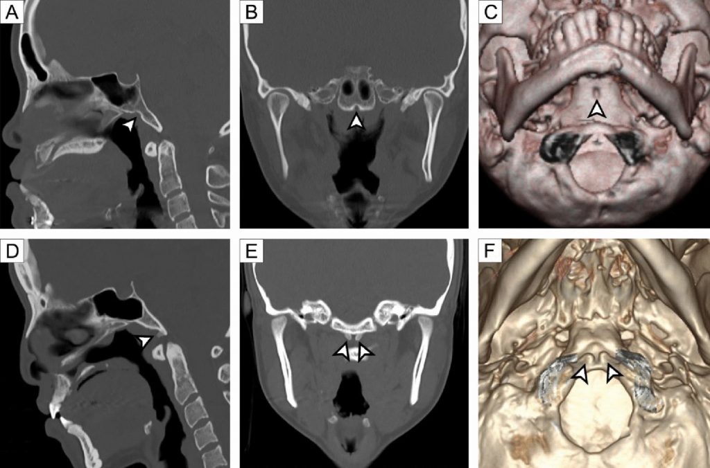
The clivus is an important landmark in neurosurgery and radiology due to its proximity to critical neurovascular structures. Anatomical variations of the clivus are diverse and may mimic pathological lesions on imaging. However, data on their prevalence in Southeast Asian populations are limited. This study investigated the prevalence and morphology of clival anatomical variations in the Thai population using computed tomography (CT). A total of 429 cranial CT scans (233 males and 196 females; mean age 39.5 ± 15.4 years) from patients without cranial abnormalities were retrospectively analyzed. The CT images were examined for fossa navicularis magna (FNM), basilar processes (BP), canalis basilaris medianus (CBM), tubercle at the anterior margin of the foramen magnum (TFM), condylus tertius (CT), and arcus praebasiooccipitalis (AP). FNM was the most common variation, identified in 107 cases (24.9%), followed by CBM (9.6%), BP (7.2%), and TFM (4.9%). CT (1.6%) and AP (0.5%) were rare. The CBM appeared as superior recess (5.1%), channel (2.6%), and inferior recess (2.3%) types. No significant sex differences were found for any variant (p > 0.05). These findings indicate that clival variations are relatively common in the Thai population, with FNM being the most prevalent. Knowledge of these variations is essential for accurate radiological interpretation and for planning neurosurgical procedures involving the skull base. Population-specific data such as these are crucial to improve diagnostic accuracy and surgical safety among Thai individuals.
KEYWORDS: clivus variation, anatomical variation, computed tomography, fossa navicularis magna, canalis basilaris medianus, skull base
Citation: Yurasakpong, L., Suwannakhan, A., Asuvapongpatana, S. et al. Radiological Assessment of Clival Morphological Variants in the Thai Population Using Computed Tomography. Bratisl. Med. J. (2025). https://doi.org/10.1007/s44411-025-00438-5
RELATED SDGs:
3. GOOD HEALTH AND WELL-BEING

ผู้ให้ข้อมูล: ผู้ช่วยศาสตราจารย์ ดร.อธิคุณ สุวรรณขันธ์
ชื่ออาจารย์ที่ทำวิจัย: ผู้ช่วยศาสตราจารย์ ดร.ลภัสรดา ยุรศักดิ์พงศ์
ผู้ช่วยศาสตราจารย์ ดร.อธิคุณ สุวรรณขันธ์ รองศาสตราจารย์ ดร.สมลักษณ์ อสุวพงษ์พัฒนา
Credit ภาพ: ผู้ช่วยศาสตราจารย์ ดร.อธิคุณ สุวรรณขันธ์
Webmaster: ว่าที่ ร.อ. นเรศ จันทรังสิกุล
Tags: anatomical variation, canalis basilaris medianus, clivus variation, computed tomography, fossa navicularis magna, Skull base