Highlight:
งานวิจัยนี้บ่งชี้การติดเชื้อของ ทีลาปีนไวรัส ในสมองและไขสันหลังปลานิล โดยระบุการแพร่กระจายโดยเทคนิคการแสดงออกทางพันธุกรรม In situ hybridyzation และพบว่าสาเหตุการติดเชื้อน่าจะผ่านการทำลายระบบไหลเวียนเลือดเข้าสู่ระบบหมุนเวียนน้ำเลี้ยงสมองและไขสันหลัง ทำให้เกิดอาการผิดปกติทางระบบประสาท
ชื่องานวิจัยภาษาไทย
การตรวจหาตำแหน่งของไวรัสทิลาเปีย ทีลาปีนไวรัส ในสมองปลานิลที่ทดลองติดเชื้อ
Abstract
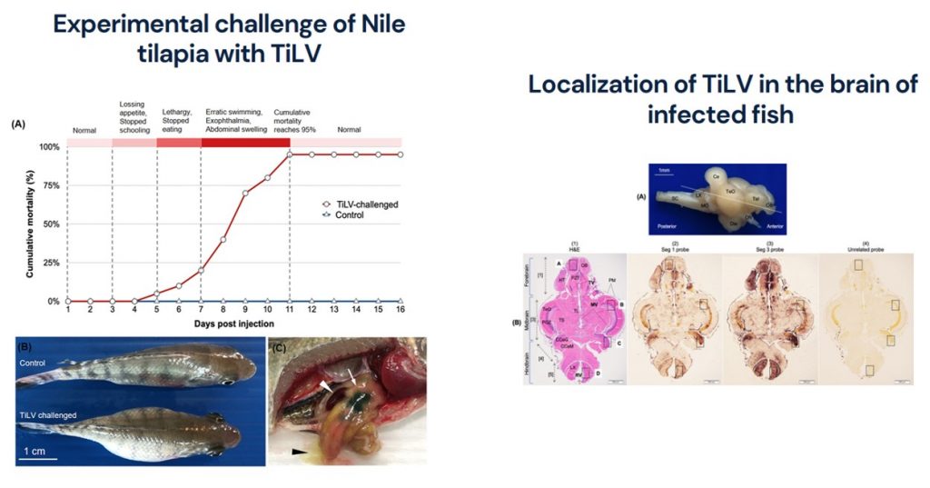
Tilapia tilapinevirus or tilapia lake virus (TiLV) is an emerging virus that inflicts significant mortality on farmed tilapia globally. Previous studies reported detection of the virus in multiple organs of the infected fish; however, little is known about the in-depth localization of the virus in the central nervous system. Herein, we determined the distribution of TiLV in the entire brain of experimentally infected Nile tilapia. In situ hybridization (ISH) using TiLV-specific probes revealed that the virus was broadly distributed throughout the brain. The strongest positive signals were dominantly detected in the forebrain (responsible for learning, appetitive behaviour and attention) and the hindbrain (involved in controlling locomotion and basal physiology). The permissive cell zones for viral infection were observed mostly to be along the blood vessels and the ventricles. This indicates that the virus may productively enter into the brain through the circulatory system and widen broad regions, possibly through the cerebrospinal fluid along the ventricles, and subsequently induce the brain dysfunction. Understanding the pattern of viral localization in the brain may help elucidate the neurological disorders of the diseased fish. This study revealed the distribution of TiLV in the whole infected brain, providing new insights into fish–virus interactions and neuropathogenesis.
ที่มาและความสำคัญ
Tilapia tilapinevirus หรือ tilapia lake virus (TiLV) เป็นไวรัสอุบัติใหม่ที่สร้างความเสียหายให้กับปลานิลที่เลี้ยงในฟาร์มทั่วโลก การศึกษาก่อนหน้านี้รายงานการตรวจพบไวรัสในหลายอวัยวะของปลาที่ติดเชื้อ อย่างไรก็ตาม ยังไม่ค่อยมีความรู้เชิงลึกถึงการกระจายตัวของไวรัสชนิดนี้ในระบบประสาทส่วนกลางของปลา งานวิจัยนี้ได้ศึกษาการกระจายตัวของไวรัส TiLV ในสมองทุกส่วนของปลานิลที่ถูกทำให้ติดเชื้อในห้องปฏิบัติการ ผลการศึกษาด้วยเทคนิค in situ hybridization โดยใช้โพรบ TiLV จำเพาะ พบไวรัสอยู่ในทุกส่วนทั่วสมอง ตำแหน่งที่ให้ผลบวกที่เด่นชัดที่สุดอยู่ในสมองส่วนหน้า (รับผิดชอบการเรียนรู้ การอยากอาหาร และความสนใจ) และสมองส่วนหลัง (เกี่ยวข้องกับการควบคุมการเคลื่อนไหวและสรีรวิทยาพื้นฐาน) บริเวณเซลล์ติดเชื้อโดยส่วนใหญ่อยู่ตามหลอดเลือดและโพรงสมอง ข้อมูลเหล่านี้บ่งชี้ว่าไวรัสอาจเข้าสู่สมองผ่านระบบไหลเวียนเลือดและขยายขอบเขตกว้างออกไป โดยอาจผ่านทางน้ำหล่อเลี้ยงสมองไปตามโพรงสมอง จนทำให้เกิดความผิดปกติของสมองในที่สุด การทำความเข้าใจรูปแบบของการกระจายตัวของไวรัสในสมองอาจช่วยอธิบายได้ถึงความผิดปกติของระบบประสาทของปลาป่วย การศึกษาครั้งนี้เผยให้เห็นการกระจายของ TiLV ในสมองปลา ซึ่งเป็นข้อมูลใหม่เชิงลึกเกี่ยวกับปฏิสัมพันธ์ระหว่างปลากับไวรัส และการเกิดโรคทางระบบประสาทของปลา
KEYWORDS: blood–brain barrier, brain, cerebrospinal fluid, in situ hybridization, localization, neuropathogenesis, tilapia lake virus, ventricles
Citation:
Dinh-Hung, N., Sangpo, P., Kruangkum, T., …Rodkhum, C., Dong, H.T. 2021. Dissecting the localization of Tilapia tilapinevirus in the brain of the experimentally infected Nile tilapia, Oreochromis niloticus (L.). Journal of Fish Diseases, 44(8), pp. 1053–1064.
https://doi.org/10.1111/jfd.13367
RELATED SDGs:
SDG Goal หลัก ที่เกี่ยวข้อง
14. LIFE BELOW WATER

SDG Goal ที่เกี่ยวข้องอื่น ๆ
2. ZERO HUNGER

ผู้ให้ข้อมูล: ผู้ช่วยศาสตราจารย์ ดร.ธนพงศ์ เครื่องคำ
ชื่ออาจารย์ที่ทำวิจัย: ผู้ช่วยศาสตราจารย์ ดร.ธนพงศ์ เครื่องคำ
ภาพถ่าย: ผู้ช่วยศาสตราจารย์ ดร.ธนพงศ์ เครื่องคำ
Tags: blood–brain barrier, brain, cerebrospinal fluid, in situ hybridization, localization, neuropathogenesis, tilapia lake virus, ventricles