Highlight
รายงานนี้นำเสนอกล้ามเนื้อเสตอร์นารีสที่มีหัวกล้ามเนื้อเจ็ดหัว ซึ่งเป็นความแปรผันทางกายวิภาคที่ไม่เคยมีการอธิบายมาก่อน พร้อมภาพสามมิติจากโฟโตแกรมเมตรี ข้อมูลนี้เพิ่มความเข้าใจเชิงลึกที่สำคัญสำหรับแพทย์และนักรังสีวิทยาในการประเมินและวางแผนรักษาทางคลินิก
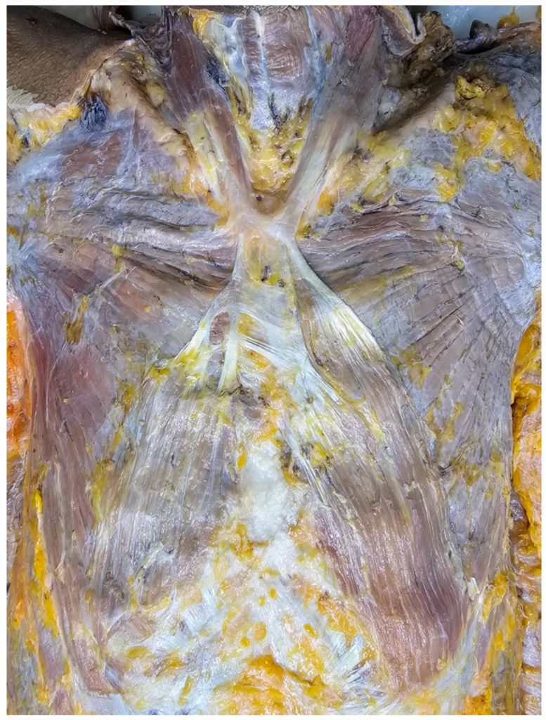
ที่มาและความสำคัญ
กล้ามเนื้อเสตอร์นารีส (sternalis muscle) เป็นกล้ามเนื้อแปรผันทางกายวิภาคที่พบได้บนผนังทรวงอก แม้ว่าจะถูกรายงานมานานในการศึกษากายวิภาค แต่ยังคงไม่คุ้นเคยสำหรับแพทย์และนักรังสีวิทยา การศึกษานี้ได้นำเสนอกรณีความแปรผันที่ไม่เคยรายงานมาก่อนของกล้ามเนื้อเสตอร์นารีสที่มีหัวกล้ามเนื้อเจ็ดหัว ในผู้บริจาคร่างกายเพศชายอายุ 79 ปี โดยหัวกล้ามเนื้อสองหัวเกิดจากข้อต่อของกล้ามเนื้อสเตอร์โนคลีโดมาโสตอยด์และห้าหัวเกิดจากเยื่อหุ้มกล้ามเนื้อเพคโตริลิสเมเจอร์ พร้อมด้วยแบบจำลองสามมิติด้วยวิธีโฟโตแกรมเมตรีที่ช่วยให้เห็นรายละเอียดของข้อความแปรผันนี้อย่างชัดเจน ข้อมูลดังกล่าวมีประโยชน์ต่อแพทย์ผู้ผ่าตัดเฉพาะทาง โดยเฉพาะในบริบทการผ่าตัดเต้านมและการตีความภาพกายวิภาคของผนังทรวงอก
Abstract
The sternalis muscle, a well-documented anatomical variation in the chest muscles, has garnered attention in anatomical research but remains relatively unfamiliar to clinicians and radiologists. This variation exhibits a wide array of descriptions and classifications in the literature, emphasizing its highly variable characteristics. This study presents a new variant of the sternalis muscle with seven muscle bellies in a 79-year-old male donor. Bilateral accessory heads of the sternocleidomastoid muscles gave rise to two superior heads. Furthermore, five additional heads originated from the pectoralis major fascia, with three on the left and two on the right, together having widths of 6.6 cm on the left and 5.3 cm on the right. Innervation of the inferior heads was provided by the intercostal nerves. The configuration of the sternalis muscle with seven heads found in this study is exceptionally distinctive and has never been reported. This unique anatomical variation, coupled with three-dimensional imaging using photogrammetry, offers valuable insights for clinicians, especially in the context of breast surgery.
KEYWORDS: sternalis muscle, anatomical variation, thoracic wall, photogrammetry, three-dimensional imaging
Citation: Berkban, T., Tangsrisakda, N., Sritawan, N., Samrid, R., Senarai, T., Taradolpisut, N., Yurasakpong, L., & Suwannakhan, A. (2025). Seven-Headed Sternalis: A Case Report with Three-Dimensional Presentation Using Photogrammetry. Diagnostics, 15(23), 3033. https://doi.org/10.3390/diagnostics15233033
RELATED SDGs:
3. GOOD HEALTH AND WELL-BEING

ผู้ให้ข้อมูล: ผู้ช่วยศาสตราจารย์ ดร.อธิคุณ สุวรรณขันธ์
ชื่ออาจารย์ที่ทำวิจัย: ผู้ช่วยศาสตราจารย์ ดร.ลภัสรดา ยุรศักดิ์พงศ์
ผู้ช่วยศาสตราจารย์ ดร.อธิคุณ สุวรรณขันธ์
Credit ภาพ: ผู้ช่วยศาสตราจารย์ ดร.อธิคุณ สุวรรณขันธ์
Webmaster: ว่าที่ ร.อ. นเรศ จันทรังสิกุล
Tags: anatomical variation, photogrammetry, sternalis muscle, thoracic wall, three-dimensional imaging