Highlight
รายงานนี้นำเสนอรูเปิดปล่อยเลือดที่ผิดปกติในกระดูกขมับของผู้ป่วยชายอายุ 64 ปี ซึ่งไม่ตรงกับรูเปิดที่อธิบายไว้ก่อนหน้านี้ การรู้จักรูปแบบความแปรผันเช่นนี้สำคัญเพื่อลดการตีความผิดพลาดในภาพถ่ายและป้องกันภาวะแทรกซ้อนในการผ่าตัดฐานกะโหลกศีรษะ
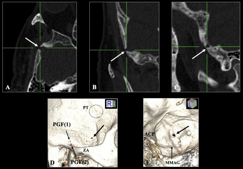
ที่มาและความสำคัญ
รูเปิดปล่อยเลือด (emissary foramen) เป็นช่องขนาดเล็กในกระดูกกะโหลกศีรษะที่เชื่อมต่อระบบหลอดเลือดดำภายในกะโหลกกับเครือข่ายหลอดเลือดดำภายนอก ทั้งนี้รูเปิดปล่อยเลือดที่พบบ่อยได้รับการอธิบายอย่างดีแล้ว แต่ความแปรผันที่ผิดปกติยังไม่ได้รับความสนใจเพียงพอ การศึกษานี้รายงานกรณีรูเปิดปล่อยเลือดที่ไม่ปกติในกระดูกข้างขมับของผู้ป่วยชายอายุ 64 ปีจากภาพถ่ายเอกซเรย์คอมพิวเตอร์ความละเอียดสูง พบช่องเปิดขนาด 3.15 มม. ที่แยกออกจากโพสท์เกลนอยด์ฟอราเมนและไม่พบในฝั่งตรงข้าม โดยตำแหน่งของรูเปิดนี้มีแนวโน้มแสดงการเชื่อมต่อหลอดเลือดดำจากพีทีรอยด์เวนัสเพล็กซัสสู่ช่องหลอดเลือดดำภายในกะโหลก การรับรู้รูเปิดที่ผิดปกติประเภทนี้มีความสำคัญต่อการตีความภาพทางการแพทย์ ป้องกันการวินิจฉัยผิดพลาด และเตรียมการผ่าตัดฐานกะโหลกศีรษะอย่างรอบคอบเพื่อหลีกเลี่ยงภาวะแทรกซ้อนเช่นเลือดออกโดยไม่คาดคิด
Abstract
The sternalis muscle, a well-documented anatomical variation in the chest muscles, has garnered attention in anatomical research but remains relatively unfamiliar to clinicians and radiologists. This variation exhibits a wide array of descriptions and classifications in the literature, emphasizing its highly variable characteristics. This study presents a new variant of the sternalis muscle with seven muscle bellies in a 79-year-old male donor. Bilateral accessory heads of the sternocleidomastoid muscles gave rise to two superior heads. Furthermore, five additional heads originated from the pectoralis major fascia, with three on the left and two on the right, together having widths of 6.6 cm on the left and 5.3 cm on the right. Innervation of the inferior heads was provided by the intercostal nerves. The configuration of the sternalis muscle with seven heads found in this study is exceptionally distinctive and has never been reported. This unique anatomical variation, coupled with three-dimensional imaging using photogrammetry, offers valuable insights for clinicians, especially in the context of breast surgery.
KEYWORDS: Emissary foramen, Temporal bone, Anatomical variation, Skull base, Venous anatomy
Citation: Triantafyllou, G., Papadopoulos-Manolarakis, P., Suwannakhan, A., & Piagkou, M. (2025). An unusual emissary foramen of the temporal bone. Surgical and Radiologic Anatomy, 47, Article 236. https://doi.org/10.1007/s00276-025-03753-2
RELATED SDGs:
3. GOOD HEALTH AND WELL-BEING

ผู้ให้ข้อมูล: ผู้ช่วยศาสตราจารย์ ดร.อธิคุณ สุวรรณขันธ์
ชื่ออาจารย์ที่ทำวิจัย: ผู้ช่วยศาสตราจารย์ ดร.อธิคุณ สุวรรณขันธ์
Credit ภาพ: ผู้ช่วยศาสตราจารย์ ดร.อธิคุณ สุวรรณขันธ์
Webmaster: ว่าที่ ร.อ. นเรศ จันทรังสิกุล
Tags: anatomical variation, Emissary foramen, Skull base, Temporal bone, Venous anatomy