Highlight:
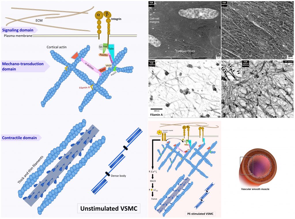
การปรับสภาพของโครงสร้างเซลล์กล้ามเนื้อเรียบที่บุผนังหลอดเลือด (vascular smooth muscle cell) แสดงลักษณะการแตกแขนงของกลุ่มเส้นใยแอคติน (cortical actin cytoskeleton) และกลุ่มโปรตีนที่เกาะ (focal adhesion proteins) ได้แก่ talin, zyxin, filamin A ในบริเวณชิดขอบเซลล์ในสภาวะปกติและเมื่อเซลล์ถูกกระตุ้นการหดตัวด้วย phenylephrine ถูกศึกษาโดยใช้เทคนิคย้อมอิมมูโนภายในเซลล์กล้ามเนื้อเรียบและเคลือบผิวเซลล์ด้วยแผ่นคาร์บอนบาง เพื่อส่องดูด้วยกล้องจุลทรรศน์อิเล็กตรอน
ที่มาและความสำคัญ
เส้นใยแอคตินมีบทบาทสำคัญต่อการหดตัวของเซลล์กล้ามเนื้อเรียบ โดยทำงานร่วมกับเส้นใยไมโอซินในการเกิดครอสบริดจ์ นอกจากนี้เส้นใยแอคตินยังส่งแรงอันมีผลต่อการหดตัวของเซลล์ต่อไปยังสารเคลือบนอกเซลล์และไปยังเซลล์ที่อยู่ข้างเคียง ปัจจัยที่มีผลต่อแรงของการหดตัวจึงถูกควบคุมเริ่มต้นมาจากเส้นใยแอคตินที่อยู่บริเวณชิดขอบเซลล์ (cortical actin filament) ซึ่งมีหน้าที่ถ่ายทอดสัญญาณเชิงกลและเชิงเคมีที่เยื่อหุ้มเซลล์ด้วย จากข้อมูลนี้จึงคาดว่าเมื่อเซลล์กล้ามเนื้อเรียบหดตัวจากปัจจัยภายในเซลล์หรือตอบสนองผ่านสิ่งเร้าเชิงกลและเชิงเคมีจากภายนอกเซลล์ เส้นใยแอคตินจะมีการปรับสภาพ (remodeling) จากการศึกษาพบว่าการทำงานของเซลล์กล้ามเนื้อเรียบที่บุผนังหลอดเลือดมีความสัมพันธ์กับการสภาพและบทบาทของกลุ่มเส้นใย (cytoskeleton) และโปรตีนที่เกี่ยวข้องภายในเซลล์ อันส่งผลต่อการปรับสภาพของเซลล์กล้ามเนื้อเรียบตามแรงยึดหรือแรงดันที่เกิดจากภายนอกเซลล์ได้ (plasticity) งานวิจัยนี้เราใช้เทคนิคย้อมอิมมูโนภายในเซลล์กล้ามเนื้อเรียบที่สามารถหดและคลายตัวได้และเคลือบผิวเซลล์ด้วยแผ่นคาร์บอนบาง ทำให้สามารถศึกษาโครงสร้างของกลุ่มเส้นใยแอคตินในบริเวณชิดขอบเซลล์รวมถึงโปรตีนที่เกาะอยู่โดยใช้กล้องจุลทรรศน์อิเล็กตรอนที่เอียงตัวอย่าง +10 และ -10 องศา เราพบว่ากลุ่มเส้นใยแอคตินที่อยู่บริเวณชิดขอบเซลล์แตกแขนงออกและเชื่อมต่อระหว่างกัน โดยมีโปรตีน ที่เกาะอยู่ในตำแหน่งเฉพาะ (focal adhesion proteins) ช่วยคงสภาพของกลุ่มเส้นใยไว้ จากการศึกษาโครงสร้างละเอียดถึงระดับนาโนเมตรทำให้สามารถติดตามโปรตีน talin และ zyxin โดย talin เป็นโปรตีนเกาะอยู่กับเส้นใยแอคตินกับ integrin บนเยื่อหุ้มเซลล์ที่จะเชื่อมต่อกับโมเลกุลอื่นด้านนอกเซลล์ ส่วน zyxin เป็นโปรตีนเกาะบนเส้นใยแอคตินเชื่อมอยู่ระหว่าง alpha-actinin และ VASP ที่มีส่วนช่วยในการทำงานของเซลล์กล้ามเนื้อเรียบ เมื่อใช้สาร phenylephrine เพื่อกระตุ้นการหดตัวของเซลล์จะพบการแตกแขนงของเส้นใยแอคตินที่บริเวณชิดขอบเซลล์ โดยไม่มีการเปลี่ยนแปลงของระยะห่างระหว่างโปรตีน talin ที่เกาะกับแขนงเส้นใยแอคติน แต่พบระยะที่สั้นลงอย่างมีนัยสำคัญของโปรตีน zyxin ที่ระหว่างแขนงเส้นใย ผลการศึกษานี้หักล้างกับข้อมูลเดิมที่เชื่อว่ากลุ่มเส้นใยแอคตินมักไม่มีการปรับสภาพเมื่อมีการหดตัวของเซลล์กล้ามเนื้อเรียบ การส่องดูโครงสร้างเส้นใยจากเซลล์โดยใช้กล้องจุลทรรศน์อิเล็กตรอนในครั้งนี้ช่วยยืนยันการปรับสภาพของกลุ่มโปรตีนที่เกาะกับเส้นใยที่ตำแหน่งจำเพาะเมื่อเซลล์กล้ามเนื้อเรียบที่บุผนังหลอดเลือดมีการหดและคลายตัวในสภาพปกติ ์
Abstract
Considerable controversy has surrounded the functional anatomy of the cytoskeleton of the contractile vascular smooth muscle cell. Recent studies have suggested a dynamic nature of the cortical cytoskeleton of these cells, but direct proof has been lacking. Here, we review past studies in this area suggesting a plasticity of smooth muscle cells. We also present images testing these suggestions by using the technique of immunoelectron microscopy of metal replicas to directly visualize the cortical actin cytoskeleton of the contractile smooth muscle cell along with interactions by representative cytoskeletal binding proteins. We find the cortical cytoskeletal matrix to be a branched, interconnected network of linear actin bundles. Here, the focal adhesion proteins talin and zyxin were localized with nanometer accuracy. Talin is reported in past studies to span the integrin–cytoplasm distance in fibroblasts and zyxin is known to be an adaptor protein between alpha-actinin and VASP. In response to activation of signal transduction with the alpha-agonist phenylephrine, we found that no movement of talin was detectable but that the zyxin-zyxin spacing was statistically significantly decreased in the smooth muscle cells examined. Contractile smooth muscle is often assumed to have a fixed cytoskeletal structure. Thus, the results included here are important in that they directly support the concept at the electron microscopic level that the focal adhesion of the contractile smooth muscle cell has a dynamic nature and that the protein–protein interfaces showing plasticity are protein-specific.
KEYWORDS: actin, focal adhesion proteins, immunoelectron microscopy, vascular smooth muscle
Citation: Suphamungmee W, Lehman W, Morgan KG. Functional Remodeling of the Contractile Smooth Muscle Cell Cortex, a Provocative Concept, Supported by Direct Visualization of Cortical Remodeling. Biology. 2022; 11(5):662.
DOI: https://doi.org/10.3390/biology11050662
RELATED SDGs:
SDG Goal หลัก ที่เกี่ยวข้อง
3. GOOD HEALTH AND WELL-BEING

ผู้ให้ข้อมูล: ผู้ช่วยศาสตราจารย์ ดร.วรวิทย์ ศุภมั่งมี
ชื่ออาจารย์ที่ทำวิจัย: ผู้ช่วยศาสตราจารย์ ดร.วรวิทย์ ศุภมั่งมี
ภาพถ่าย: ผู้ช่วยศาสตราจารย์ ดร.วรวิทย์ ศุภมั่งมี
Tags: actin, focal adhesion proteins, immunoelectron microscopy, vascular smooth muscle