Highlight
การรักษาแผลที่ข้อเท้าโดยการปลูกถ่ายผิวหนังและเนื้อเยื่อจากหลังเท้าในอาจารย์ใหญ่แบบนิ่มเสมือนจริง

ที่มาและความสำคัญ
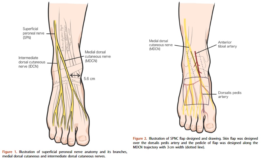
บาดแผลบริเวณข้อเท้าที่ตื้นและเล็กสามารถรักษาโดยการเย็บปิดแผลปกติ แต่บาดแผลที่ลึกจนถึงเส้นเอ็นหรือกระดูกและกว้างเกินกว่าที่จะเย็บปิดควรรักษาด้วยการปลูกถ่ายผิวหนังและเนื้อเยื่อจากบริเวณใกล้เคียงกับแผลซึ่งจะให้ผลการรักษาที่ดีกว่า ผู้วิจัยได้ศึกษาผิวหนังและเนื้อเยื่อบริเวณหลังเท้าซึ่งถูกเลี้ยงโดยแขนงเส้นประสาทและแขนงหลอดเลือดจำเพาะเพื่อปลูกถ่ายทดแทนบริเวณแผลที่ข้อเท้าในร่างอาจารย์ใหญ่แบบนิ่มเสมือนจริง ข้อดีของการใช้ผิวหนังและเนื้อเยื่อบริเวณหลังเท้าเพื่อรักษาแผลที่ข้อเท้า ได้แก่ เนื้อเยื่อหลังเท้ามีขนาดบางเพราะไขมันใต้ผิวหนังน้อยทำให้หลังปลูกถ่ายที่ข้อเท้าแล้วไม่ขัดขวางการเคลื่อนไหวของข้อเท้าและการสวมใส่รองเท้า ทั้งเนื้อเยื่อที่ปลูกถ่ายมีเส้นประสาทเลี้ยงจึงทำให้ผิวหนังบริเวณที่ปลูกถ่ายทดแทนสามารถรับความรู้สึกได้ และเนื้อเยื่อและกล้ามเนื้อที่หลังเท้า ณ ตำแหน่งเดิมยังเป็นปกติเนื่องจากหลอดเลือดหลักที่เลี้ยงเท้ายังคงอยู่

Abstract
Background
Soft tissue defects around the ankle are common and must be covered with thin and pliable flaps. A regional flap, particularly from the dorsum of the foot was considered ideal. A neurocutaneous flap, based on the superficial peroneal nerve (SPN) and its branches was designed as a proximally based flap via cadaveric dissection. This study aimed to demonstrate the vascularity and characteristics of the superficial peroneal neurocutaneous (SPNC) flap.
Materials and Methods
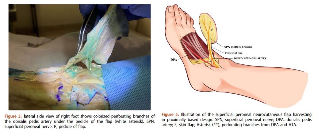
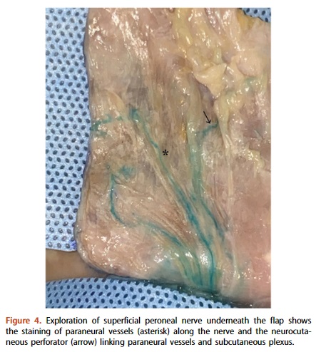
The SPNC flap was created in 11 lower limbs (seven cadavers) using a proximally based design. The skin flap was dissected at the dorsum of the foot, followed by injection of diluted methylene blue through the anterior tibial artery, to visualize the vascularity. The flap pedicle above the anterior ankle joint line was dissected along the SPN for anatomical study of perforating branches, paraneural vessels, and flap territory.
Results
The mean distances of the most proximal perforating branches were 1.51 ±1.48 cm from the anterior ankle joint line, and 5.12 ±1.78 cm from the lateral malleolus. The mean distances of the most distal perforating branches were 2.75 ± 1.54 cm from the anterior ankle joint line, and 5.90 ±1.81 cm from the lateral malleolus. The mean number of perforating branches was 3.73 ± 1.49. The mean flap territories were 5.51 ±0.59 cm in length, and 7.15 ± 0.64 cm in width.
Conclusion
The SPNC flap is an alternative method for soft tissue reconstruction around the ankle with a proximally based flap design. The antegrade flow has been shown to offer effective vascularity in flaps prepared via cadaveric dissection.

KEYWORDS: superficial peroneal neurocutaneous flap, regional flap, soft cadaver, anatomy, superficial peroneal nerve, ankle, foot
Citation: Kanchanathepsak T, Kunsook K, Panoinont W, Suriyonplengsaeng C, Suppaphol S, Watcharananan I, Tuntiyatorn P, Tawonsawatruk T. The superficial peroneal neurocutaneous flap: a cadaveric study. J Plast Surg Hand Surg. 2023 Feb-Dec;57(1-6):500-504. doi: 10.1080/2000656X.2023.2168273. Epub 2023 Jan 20. PMID: 36661749.
RELATED SDGs:
3. GOOD HEALTH AND WELL-BEING

ผู้ให้ข้อมูล: ผู้ช่วยศาสตราจารย์ นพ.ชินวุฒิ สุริยนเปล่งแสง
ชื่ออาจารย์ที่ทำวิจัย: ผู้ช่วยศาสตราจารย์ นพ.ชินวุฒิ สุริยนเปล่งแสง
แหล่งทุนวิจัย: ทุน Biomedical research จากคณะแพทยศาสตร์โรงพยาบาลรามาธิบดี
Credit ภาพ: ผู้ช่วยศาสตราจารย์ นพ.ชินวุฒิ สุริยนเปล่งแสง
Tags: Anatomy, ankle, foot, regional flap, soft cadaver, superficial peroneal nerve, superficial peroneal neurocutaneous flap
