Highlight
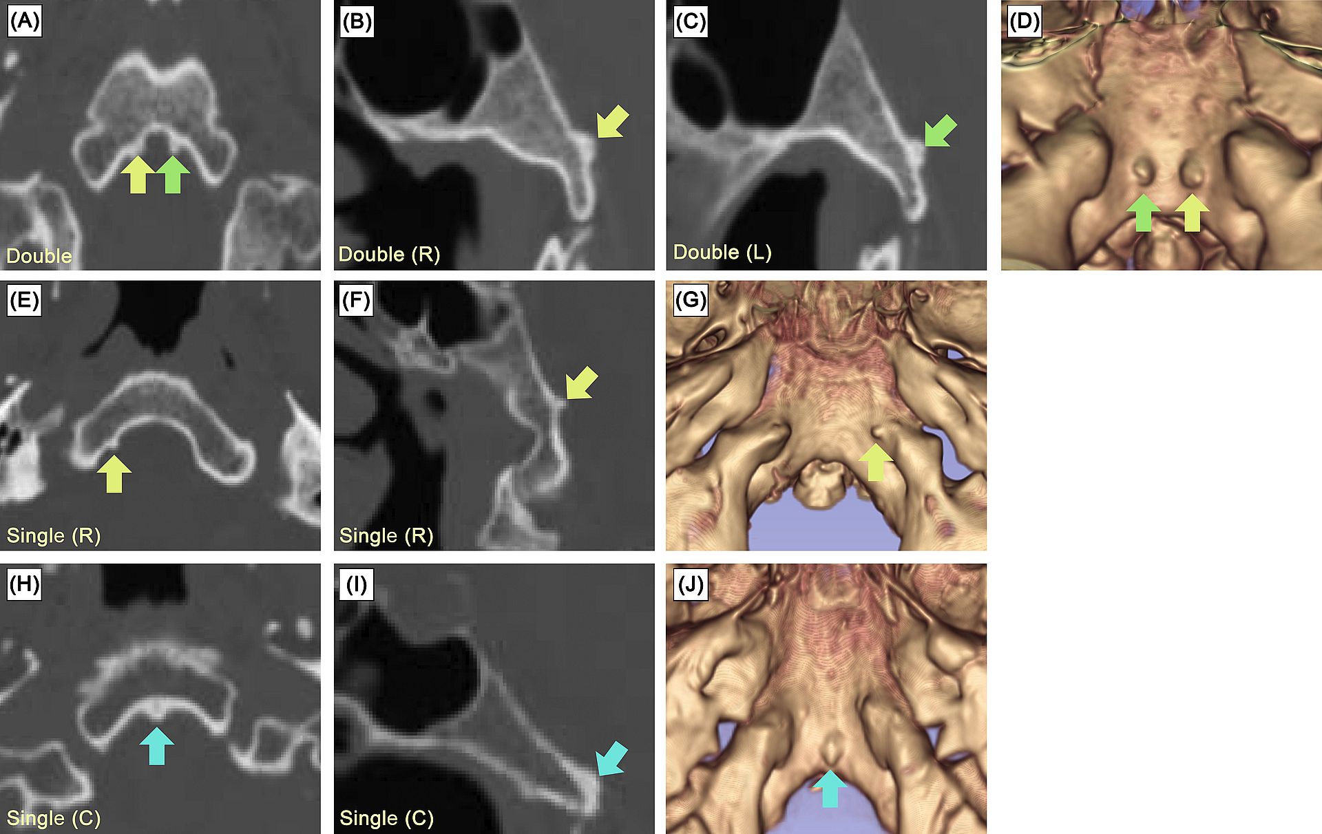
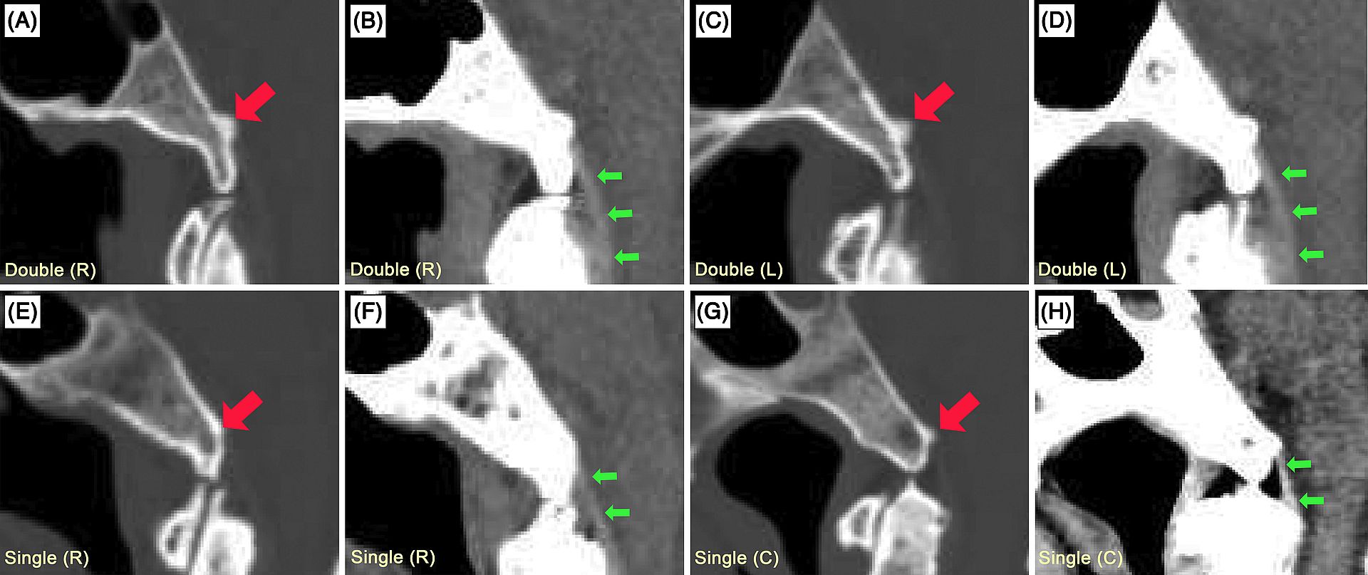
จากการศึกษาจากภาพเอกซเรย์คอมพิวเตอร์ (computed tomography) จากผู้ป่วยรวม 407 ราย ผู้วิจัยพบปุ่มกระดูกที่ยังไม่มีการอธิบายมาก่อนในส่วนบนของกระดูก clivus ในผู้ป่วย 40 รายหรือร้อยละ 9.8 โดยปุ่มกระดูกนี้วางตัวอยู่ระว่าง dorsum sallae และ foramen magnum ปุ่มกระดูกชนิดใหม่นี้สามารถจำแนกได้เป็นสามประเภท ได้แก่ single double และ triple โดยชนิด single มีความชุกที่ร้อยละ 8.7 ชนิด double มีความชุกที่ 0.98 และชนิด triple ที่ร้อยละ 0.25 ความกว้างและความสูงเฉลี่ยของปุ่มกระดูกนี้อยู่ที่ 4.4±1.5 มม. (ช่วง 1.4–7.9 มม.) และ 1.7±0.7 มม. (ช่วง 0.8–4.2 มม.) ตามลำดับ ผู้วิจัยได้บัญญัติชื่อของปุ่มกระดูกเหล่านี้ว่า “basilar tubercles of the clivus” และ “basilar eminences of the clivus” ขึ้นอยู่กับขนาดของปุ่มที่พบ


ที่มาและความสำคัญ
งานวิจัยฉบับนี้รายงานถึงการค้นพบของปุ่มกระดูกชนิดใหม่ตรงบริเวณส่วนบนของ clivus ซึ่งเป็นพื้นที่ที่อยู่ตรงฐานกะโหลกด้านหน้าต่อ foramen magnum
Abstract
Background
The clivus forms the central skull base between the dorsum sellae and the foramen magnum. Although bony variations of the inferior surface of the clivus are well-recognized and have been well studied, studies of bony variations of the basilar (superior) surface of the clivus are scarce. Therefore, the present study was performed to investigate bony anatomical variations on the basilar part of the clivus.
Methods
Computed tomography scans belonging to 407 Indian subjects from the CQ500 open-access dataset were retrospectively reviewed.
Results
Bony tubercles on the basilar surface of the clivus were found in 40 cases (9.83%). They were classified into three types including single, double and triple. A single tubercle was found in 35 cases (8.60%) including 12 on the left (2.95%), 10 on the right (2.46%) and 13 in the center (3.19%). The tubercles were doubled in four cases (0.98%) and tripled in one case (0.25%). The average width and height of the tubercles were 4.4 ± 1.5 mm (range 1.4–7.9 mm) and 1.7 ± 0.7 mm (range 0.8–4.2 mm), respectively. Ninety-five (95%) percent of the tubercles were located on the lower half of the clivus.
Conclusions
To our knowledge, these tubercles have not been previously described. Therefore, we suggest the terms “basilar tubercles of the clivus” and “basilar eminences of the clivus”, depending on their sizes. Knowledge of these newly described structures is important when interpreting radiological images of the skull base.
KEYWORDS: Clivus, Skull, Anatomical variation, Computed tomography, Terminology, Imaging
Citation: Tangrodchanapong, T., Yurasakpong, L., Suwannakhan, A., Chaiyamoon, A., Iwanaga, J., & Tubbs, R. S. (2023). Basilar tubercles and eminences of the clivus: Novel anatomical entities. Annals of Anatomy-Anatomischer Anzeiger, 250, 152133. DOI: https://doi.org/10.1016/j.aanat.2023.152133.
RELATED SDGs:
3. GOOD HEALTH AND WELL-BEING

ผู้ให้ข้อมูล: ผู้ช่วยศาสตราจารย์ ดร.อธิคุณ สุวรรณขันธ์
ชื่ออาจารย์ที่ทำวิจัย: ผู้ช่วยศาสตราจารย์ ดร.อธิคุณ สุวรรณขันธ์, อาจารย์ ดร.ลภัสรดา ยุรศักดิ์พงศ์
Credit ภาพ: ผู้ช่วยศาสตราจารย์ ดร.อธิคุณ สุวรรณขันธ์
Webmaster: ว่าที่ ร.อ. นเรศ จันทรังสิกุล
Tags: anatomical variation, Clivus, computed tomography, Imaging, skull, Terminology
