Highlight:
พัฒนาการของหัวใจที่สามารถติดตามได้ด้วยตัวเองผ่านภาพสามมิติที่สามารถหมุนดูเพื่อศึกษาพร้อมรายละเอียดพัฒนาการของหัวใจทั้งเชิงโครงสร้างและเชิงการปริมาณเปรียบเทียบ นำไปสู่พลวัตพัฒนาการของหัวใจที่ชัดเจนมากขึ้นกว่าในตำราทั่วไป
ที่มาและความสำคัญ
หัวใจเป็นโครงสร้างที่มีความซับซ้อนในการพัฒนาระหว่างอยู่ในครรภ์เป็นอย่างมากในตัวอ่อนมนุษย์ เนื่องจากพัฒนาการของหัวใจมีความเป็นสามมิติสูงมาก จากท่อตรง ๆ เกิดการขดงอต่าง ๆ จนได้ออกมาเป็นรูปร่างที่พบในมนุษย์ อีกทั้งโครงสร้างภายในเองยังมีการเจริญที่ต้องมีการสอดประสานกันอย่างเหมาะสมเพื่อให้ห้องตัวใจและหลอดเลือดเข้าและออกจากหัวใจทำหน้าที่ได้อย่างสมบูรณ์เมื่อแรกคลอด ดังนั้นการเข้าใจกลไกพัฒนาการที่ชัดเจนมากขึ้นนำไปสู่การต่อยอดทางการแพทย์ในการอธิบายและรักษาพัฒนาการที่ผิดปกติของหัวใจในตัวอ่อนมนุษย์ได้ดียิ่งขึ้น
Abstract
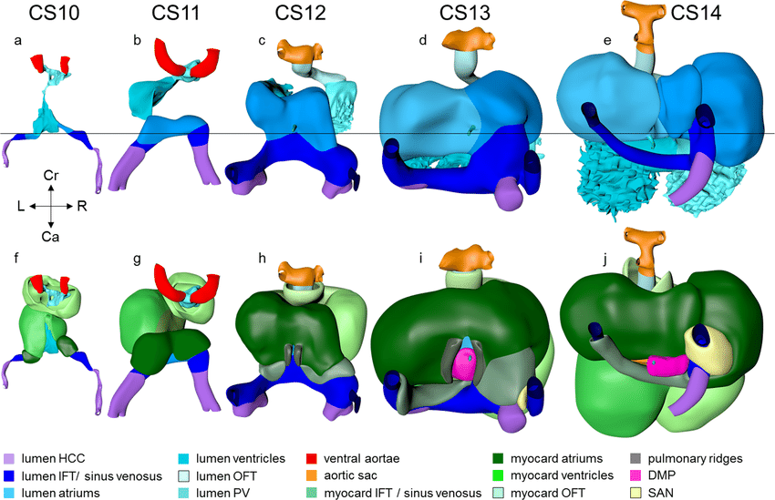
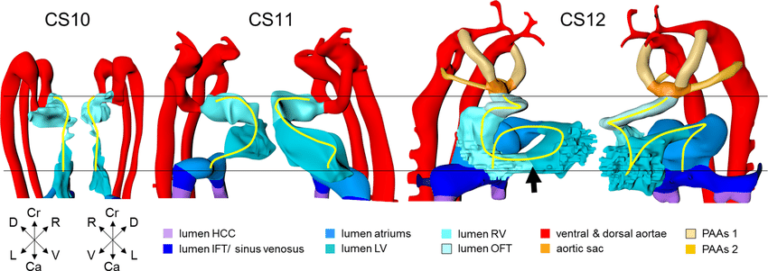
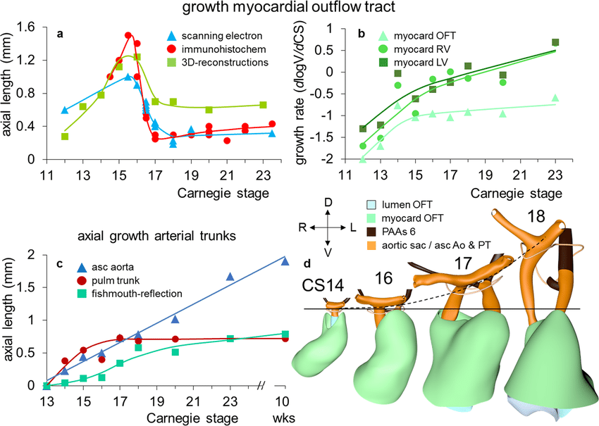
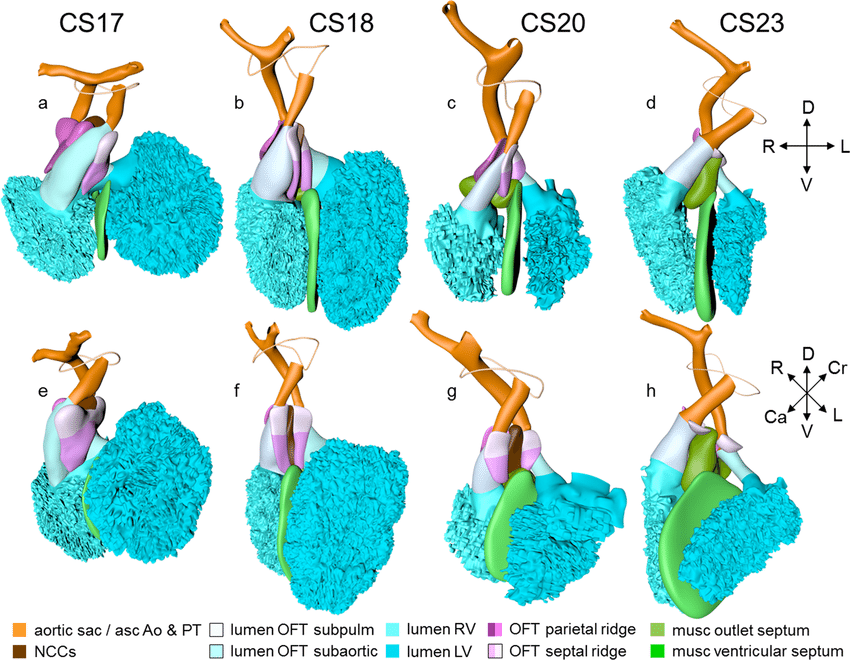
Heart development is topographically complex and requires visualization to understand its progression. No comprehensive 3-dimensional primer of human cardiac development is currently available. We prepared detailed reconstructions of 12 hearts between 3.5 and 8 weeks post fertilization, using Amira® 3D-reconstruction and Cinema4D®-remodeling software. The models were visualized as calibrated interactive 3D-PDFs. We describe the developmental appearance and subsequent remodeling of 70 different structures incrementally, using sequential segmental analysis. Pictorial timelines of structures highlight age-dependent events, while graphs visualize growth and spiraling of the wall of the heart tube. The basic cardiac layout is established between 3.5 and 4.5 weeks. Septation at the venous pole is completed at 6 weeks. Between 5.5 and 6.5 weeks, as the outflow tract becomes incorporated in the ventricles, the spiraling course of its subaortic and subpulmonary channels is transferred to the intrapericardial arterial trunks. The remodeling of the interventricular foramen is complete at 7 weeks.
KEYWORDS
heart development, 3D reconstruction, developmental dynamics
Citation
Hikspoors, J.P.J.M., Kruepunga, N., Mommen, G.M.C. et al. A pictorial account of the human embryonic heart between 3.5 and 8 weeks of development. Commun Biol5, 226 (2022).
https://doi.org/10.1038/s42003-022-03153-x
RELATED SDGs:
SDG Goal หลัก ที่เกี่ยวข้อง
4. QUALITY EDUCATION

SDG Goal ที่เกี่ยวข้องอื่น ๆ
3. GOOD HEALTH AND WELL-BEING

ผู้ให้ข้อมูล: อาจารย์ ดร.ณัฐเมธี เครือภูงา
ชื่ออาจารย์ที่ทำวิจัย: อาจารย์ ดร.ณัฐเมธี เครือภูงา
ภาพถ่าย: อาจารย์ ดร.ณัฐเมธี เครือภูงา
Tags: 3D reconstruction, developmental dynamics, heart development