Highlight
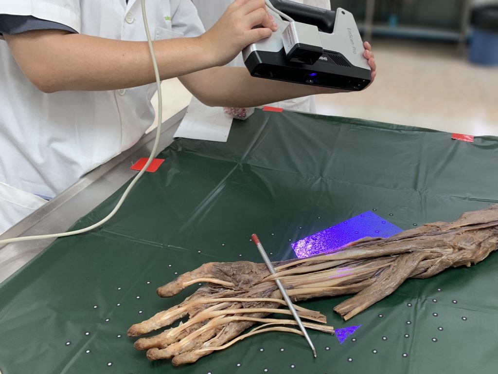
งานวิจัยฉบับนี้สแกนร่างอาจารย์ใหญ่ด้วยเครื่องแสกนแบบสามมิติและนำเสนอผลงานในรูปแบบ 3D-PDF การนำเสนอผลงานในรูปแบบนี้เป็นมิติใหม่ของการนำเสนอผลงานวิจัยและอาจเป็นมาตรฐานใหม่ในงานวิจัยทางกายวิภาคศาสตร์ในอนาคต
ที่มาและความสำคัญ
โครงสร้างแปรผัน Linburg-Comstock คือเส้นเอ็น เนื้อเยื่อเกี่ยวพัน หรือกล้ามเนื้อที่เกาะจากเส้นเอ็น flexor pollicis longus (FPL) ไปยัง flexor digitorum profundus (FDP) ของนิ้วชี้ ซึ่งพบได้ในร้อยละ 21 ของประชากรทั่วไป ซึ่ง distal interphalangeal joint ของนิ้วชี้บุคคลที่มีโครงสร้างนี้จะงอไปพร้อม ๆ กันเมื่อเกิดการหดตัวของกล้ามเนื้อ FPLงานวิจัยฉบับนี้รายงานการอุบัติขึ้นของเส้นเอ็นที่เกาะจาก FDP ไปยัง FPL ในร่างอาจารย์ใหญ่ซึ่งมีลักษณะเหมือนกับโครงสร้างแปรผัน Linburg-Comstock แบบกลับด้าน เมื่อเกิดการหดตัวของกล้ามเนื้อ FDP จะก่อให้เกิดการงอของข้อต่อ interphalangeal joint ของนิ้วโป้ง ซึ่งเห็นได้ชัดเจนเมื่อดึงเส้นเอ็นนี้ (supplementary video) นอกจากนี้ งานวิจัยฉบับนี้ได้อธิบายวิธีการแสกนตัวอย่างชิ้นส่วนจากร่างอาจารย์ใหญ่ด้วยเครื่องสแกนสามมิติแบบ fixed scan การปรับแต่งโมเดลที่ได้และการสร้างไฟล์ 3D-PDF เพื่อนำเสนอผลงานในรูปแบบสามมิติเป็นครั้งแรก
Abstract
Purpose
Two most common variations of flexor pollicis longus include its accessory head and its connection with the flexor digitorum profundus of the index (Linburg–Comstock variation). In addition, while three-dimensional (3D) screening has widely been used in anatomical education, its use as reporting tool in anatomical research is still limited. The objective of this study is to report a previously unrecognized form of the accessory head of flexor pollicis longus, discuss the potential etiology of Linburg–Comstock variation, and pilot the 3D scanning of a large-scale anatomical structure.
Methods
An unusual tendon slip was discovered during a routine dissection in the anterior compartment of the right forearm of a 54-year-old male cadaver. A 3D scanner was used to capture the surface topography of the specimen and an interactive portable document format (PDF) was created.
Results
An anomalous tendon was found originating from the lateral aspect of the flexor digitorum profundus muscle. This variant tendon then inserted onto the medial surface of the flexor pollicis longus tendon before entering the carpal tunnel. The variation resembles a reverse form of Linburg–Comstock variation, because pulling this variant tendon resulted in simultaneous flexion of the interphalangeal joint of thumb.
Conclusion
Surgeons should be aware of the reverse Linburg–Comstock variation, because it may not be detectable by the conventional provocative testing. Linburg–Comstock variation may be classified as an anatomical variant or a secondarily acquired condition depending on its type. Our demonstration of interactive 3D-PDF file highlights its potential use for delivering anatomical information in future cadaveric studies.
KEYWORDS: Flexor pollicis longus, Flexor digitorum profundus, Linburg–Comstock variation, Anatomical variations, 3D scanning, 3D visualization, Surface rendering
Citation: Prasatkaew, W., Kruepunga, N., Yurasakpong, L. et al. A reverse form of Linburg–Comstock variation with comments on its etiology and demonstration of interactive 3D portable document format. Surg Radiol Anat44, 227–232 (2022). https://doi.org/10.1007/s00276-021-02858-8
RELATED SDGs:
3. GOOD HEALTH AND WELL-BEING

ผู้ให้ข้อมูล: ผู้ช่วยศาสตราจารย์ ดร.อธิคุณ สุวรรณขันธ์
ชื่ออาจารย์ที่ทำวิจัย: ผู้ช่วยศาสตราจารย์ ดร.ณัฐเมธี เครือภูงา, ผู้ช่วยศาสตราจารย์ ดร.อธิคุณ สุวรรณขันธ์
Credit ภาพ: ผู้ช่วยศาสตราจารย์ ดร.อธิคุณ สุวรรณขันธ์
Tags: 3D scanning, 3D visualization, Anatomical variations, Flexor digitorum profundus, Flexor pollicis longus, Linburg–Comstock variation, Surface rendering