Highlight
การศึกษานี้พบความสัมพันธ์ทางกายวิภาคใหม่ระหว่างกล้ามเนื้อ anconeus epitrochlearis กับเส้นประสาทอัลนาร์ โดยพบการเชื่อมต่อโดยตรงกับ epineurium ของเส้นประสาทและแคปซูลข้อศอก กล้ามเนื้อดังกล่าวอาจมีบทบาทช่วยเคลื่อนเส้นประสาทออกจากร่องอัลนาร์ระหว่างการเคลื่อนไหวของข้อศอก มากกว่าการเป็นสาเหตุของการกดทับเส้นประสาทเพียงอย่างเดียว ผลลัพธ์มีความสำคัญต่อการวินิจฉัยและการผ่าตัดรักษา cubital tunnel syndrome
ที่มาและความสำคัญ
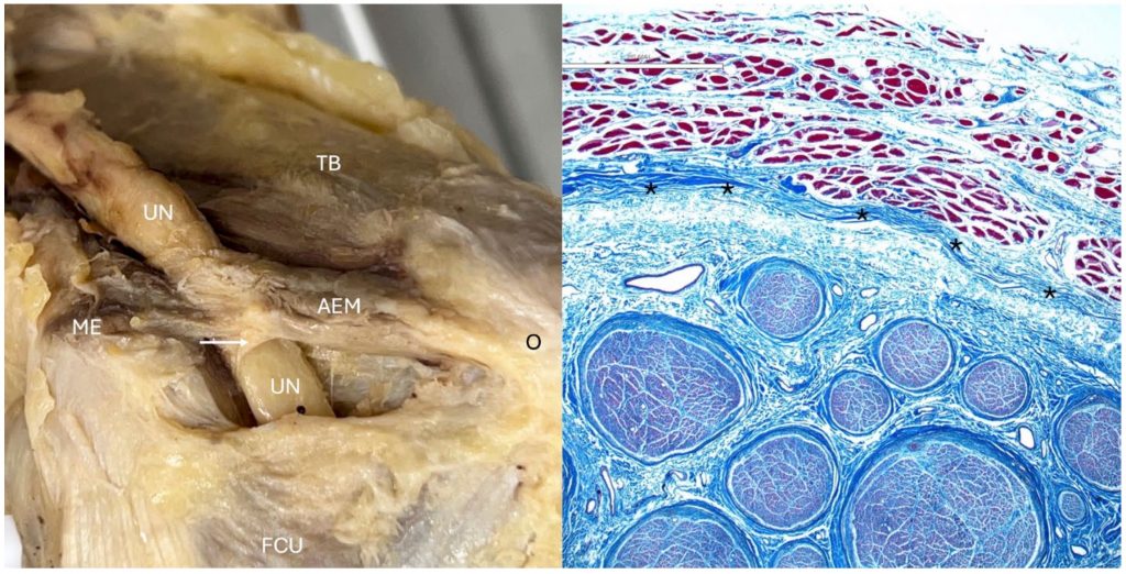
กล้ามเนื้อ anconeus epitrochlearis (AEM) เป็นความแปรผันทางกายวิภาคบริเวณข้อศอกที่พาดจาก medial epicondyle ไปยัง olecranon และเป็นส่วนหนึ่งของหลังคา cubital tunnel ซึ่งเป็นตำแหน่งที่เส้นประสาทอัลนาร์ (ulnar nerve) มักถูกกดทับจนเกิด cubital tunnel syndrome อย่างไรก็ตาม บทบาทที่แท้จริงของ AEM ต่อการเกิดหรือป้องกันภาวะดังกล่าวยังคงเป็นที่ถกเถียงกันในวรรณกรรมทางการแพทย์ บางการศึกษารายงานว่า AEM เป็นสาเหตุของการกดทับเส้นประสาท ขณะที่หลักฐานล่าสุดกลับชี้ว่าอาจมีบทบาทในการปกป้องเส้นประสาทอัลนาร์ งานวิจัยนี้จึงมุ่งศึกษาความสัมพันธ์ทางกายวิภาคระหว่าง AEM เส้นประสาทอัลนาร์ และโครงสร้างโดยรอบในศพอาจารย์ใหญ่จำนวนมาก เพื่อทำความเข้าใจกลไกที่เกี่ยวข้องกับภาวะกดทับเส้นประสาทให้ชัดเจนยิ่งขึ้น ผลการศึกษาพบความสัมพันธ์ทางกายวิภาคที่ไม่เคยมีรายงานมาก่อน โดยเฉพาะการเชื่อมต่อของ AEM กับ epineurium ของเส้นประสาทอัลนาร์และกับข้อศอก ซึ่งช่วยอธิบายบทบาทเชิงหน้าที่ของกล้ามเนื้อนี้ และมีความสำคัญต่อการวินิจฉัย การผ่าตัด และการป้องกันการบาดเจ็บของเส้นประสาทในบริเวณ cubital tunnel
Abstract
Ulnar neuropathy due to compression at the cubital tunnel is common. However, our understanding of the relationships between this type of nerve compression and the variant anconeus epitrochlearis muscle (AEM) is poorly understood. Therefore, the present anatomical study was performed to better elucidate these relationships. In 162 adult cadavers (324 sides), the roof of the cubital tunnel was dissected. The prevalence and gross anatomy of the AEM were documented. Histological and microCT analyses were performed on selected specimens to evaluate the microanatomy and radiological findings in relation to the underlying nerve, soft tissues, and bone. Additionally, with the range of motion of the elbow and artificial contraction of the AEM, the effects of the AEM on the ulnar nerve were observed. AEMs were identified on 32 (10%) of the sides. Histologically, a connective tissue connection between the AEM and ulnar nerve was found in all specimens, and a direct connection between the AEM and the underlying joint capsule was found in most specimens. No grossly visible compression of the ulnar nerve by the overlying AEM was observed with flexion or extension of the elbow. With artificial contraction of the AEM, the ulnar nerve was found to move out of the depths of the ulnar groove in roughly half of the sides. Previously unreported relationships between Osborne’s ligament, the AEM, and the underlying ulnar nerve were found. These findings will improve our understanding of the relationship between these structures and the ulnar nerve at the elbow. During surgery in this area, care should be taken to avoid injuring the ulnar nerve when applying traction to the AEM.
KEYWORDS: anatomy, cubital tunnel syndrome, elbow, nerve compression, peripheral nerve, surgery
Citation: Banerjee, S., Nguyen, K., Bishop, J. L., Pujol, J. E., Rosbrugh, J. E., Yurasakpong, L., Chaiyamoon, A., Suwannakhan, A., Dumont, A. S., Georgiev, G. P., Iwanaga, J., & Tubbs, R. S. (2026). New insights into the anconeus epitrochlearis muscle and its relationship to the ulnar nerve: Anatomical study. Clinical Anatomy, 0, 1–5. https://doi.org/10.1002/ca.70123
RELATED SDGs:
3. GOOD HEALTH AND WELL-BEING

ผู้ให้ข้อมูล: ผู้ช่วยศาสตราจารย์ ดร.อธิคุณ สุวรรณขันธ์
ชื่ออาจารย์ที่ทำวิจัย: ผู้ช่วยศาสตราจารย์ ดร.ลภัสรดา ยุรศักดิ์พงศ์ ผู้ช่วยศาสตราจารย์ ดร.อธิคุณ สุวรรณขันธ์
Credit ภาพ: ผู้ช่วยศาสตราจารย์ ดร.ลภัสรดา ยุรศักดิ์พงศ์
Webmaster: ว่าที่ ร.อ. นเรศ จันทรังสิกุล
Tags: Anatomy, cubital tunnel syndrome, Elbow, nerve compression, peripheral nerve, surgery