
Highlight
การใช้ Ultrasound elastography (UE) ถือเป็นเทคนิคใหม่ในการประเมินความยืดหยุ่นหรือความแข็งของเนื้อเยื่อที่สนใจ ดังนั้นการศึกษานี้มีวัตถุประสงค์เพื่อเปรียบเทียบความเร็วของคลื่นเฉือนในกล้ามเนื้อมดลูก (myometrium) ของมดลูกปกติ เนื้องอกในมดลูกแบบ uterine fibroid และ เนื้องอกในมดลูกแบบ adenomyosis รวมถึงประเมินความถูกต้องของการใช้ Ultrasound elastography ในการวินิจฉัยแยก adenomyosis
ที่มาและความสำคัญ
เนื้องอกมดลูกแบบ uterine fibroid และแบบ adenomyosis ถือเป็นปัญหาทางนรีเวชที่พบได้ทั่วไป แต่เนื่องจากทั้งสองภาวะทำให้เกิดอาการทางคลินิกที่คล้ายคลึงกัน ได้แก่ มดลูกผิดปกติ เลือดออกผิดปกติ การปวดในอุ้งเชิงกราน และภาวะมีบุตรยาก ดังนั้นการวินิจฉัยแยกภาวะเนื้องอกมดลูกระหว่าง uterine fibroid และ adenomyosis บางครั้งทำได้ยาก การวินิจฉัยที่แม่นยำถือเป็นสิ่งจำเป็นสำหรับสตรีที่มีภาวะมีบุตรยาก เนื่องจากทั้งสองภาวะมีทางเลือกในการรักษาที่แตกต่างกัน โดยทั่วไปการตรวจเพื่อวินิจฉัยแยกภาวะทั้งสองนี้ส่วนใหญ่จะใช้การอัลตร้าซาวด์ในการตรวจ แต่วิธีนี้มีความแม่นยำที่หลากหลายขึ้นกับประสบการณ์ของผู้ใช้ เพราะฉะนั้นการหาเครื่องมืออื่นที่มีความแม่นยำมากกว่ามาช่วยในการวินิจฉัยแยกสองภาวะนี้จึงมีความจำเป็นอย่างยิ่ง การใช้ Ultrasound elastography (UE) ถือเป็นเทคนิคใหม่ในการประเมินความยืดหยุ่นหรือความแข็งของเนื้อเยื่อที่สนใจ ดังนั้นการศึกษานี้มีวัตถุประสงค์เพื่อเปรียบเทียบความเร็วของคลื่นเฉือนในกล้ามเนื้อมดลูก (myometrium) ของมดลูกปกติ เนื้องอกในมดลูกแบบ uterine fibroid และ เนื้องอกในมดลูกแบบ adenomyosis รวมถึงประเมินความถูกต้องของการใช้ Ultrasound elastography ในการวินิจฉัยแยก adenomyosis
Abstract
Background
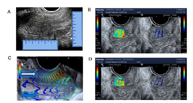
The differential diagnosis between uterine fibroid and adenomyosis is sometimes difficult; a precise diagnosis is required in women with infertility because of the different choice of treatments. Ultrasound elastography (UE) is a novel technique to evaluate the elasticity or the stiffness of the tissue of interest. The present study aims to compare UE shear wave velocity (SWV) among normal uterine myometrium, uterine fibroid, and adenomyosis, and assess the accuracy of shear wave elastography in the diagnosis of adenomyosis.
Materials and Methods
This cross-sectional study recruited 25 subjects for each group (control, adenomyosis, and fibroid) from April 2019 to April 2020. Transvaginal UE using an Aplio 500 (Toshiba Medical Systems, Japan) with ultrasound mapping for point of tissue biopsy was performed for all subjects. The diagnosis was confirmed by histology. Masson’s trichrome staining for collagen was performed and quantified
Results
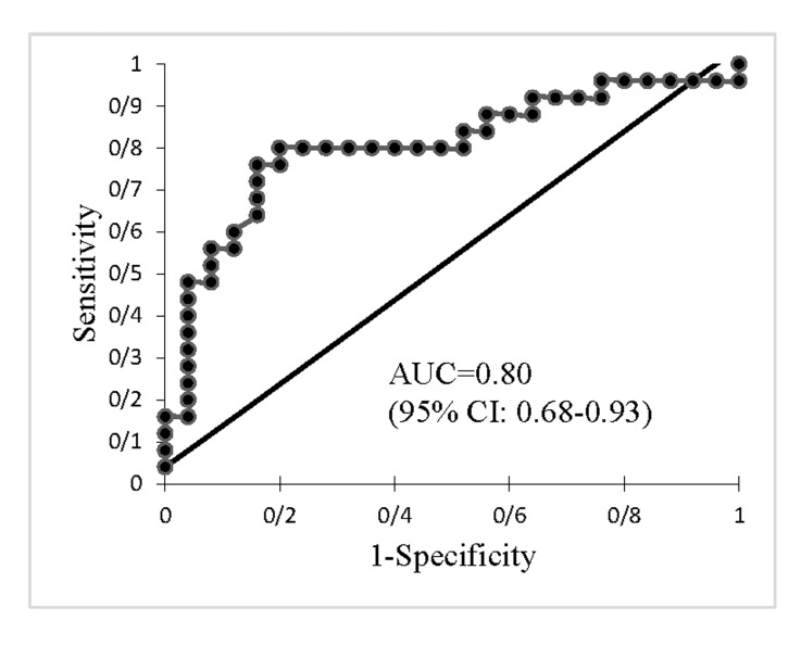
The mean ± standard deviation (SD) for SWV was 3.44 ± 0.95 m/seconds (control group), 4.63 ± 1.45 m/seconds (adenomyosis group), and 4.53 ± 1.07 m/seconds (fibroid group). The mean SWV differed when comparing normal myometrium and adenomyosis after adjustments for age and endometrial pathology (P=0.019). The cut-off point of SWV at 3.465 m/seconds could differentiate adenomyosis from the normal uterus with an 80% sensitivity, 80% specificity, and an area under the curve (AUC) of 0.80 (95% confidence interval [CI]: 0.68-0.93) (P<0.001). No significant difference in SWV between the adenomyosis and fibroid groups was detected.
Conclusion
Shear wave elastography could be an alternative tool to distinguish between normal myometrium and adenomyosis; however, it could not differentiate adenomyosis from uterine fibroid or uterine fibroid from normal myometrium.
KEYWORDS: Adenomyosis, Elasticity, Elasticity Imaging Techniques, Leiomyoma, Uterus
Citation: Pongpunprut S, Panburana P, Wibulpolprasert P, Waiyaput W, Sroyraya M, Chansoon T, Sophonsritsuk A. A Comparison of Shear Wave Elastography between Normal Myometrium, Uterine Fibroids, and Adenomyosis: A Cross-Sectional Study. Int J Fertil Steril. 2022 Jan;16(1):49-54. doi: 10.22074/IJFS.2021.523075.1074. PMID: 35103432; PMCID: PMC8808257.
RELATED SDGs:
SDG Goal หลัก ที่เกี่ยวข้อง
3. GOOD HEALTH AND WELL-BEING

ผู้ให้ข้อมูล: ผู้ช่วยศาสตราจารย์ ดร.มรกต สร้อยระย้า
ชื่ออาจารย์ที่ทำวิจัย: ผู้ช่วยศาสตราจารย์ ดร.มรกต สร้อยระย้า
Tags: Adenomyosis, Elasticity, Elasticity Imaging Techniques, Leiomyoma, Uterus
