
ชื่องานวิจัยภาษาไทย
การประเมินประโยชน์ทางการวินิจฉัยของตัวบ่งชี้การอักเสบในชิ้นกล้ามเนื้อลายซึ่งรักษาสภาพด้วยฟอร์มาลินและฝังพาราฟินสำหรับการวินิจฉัยโรคกล้ามเนื้ออักเสบชนิดไม่ทราบสาเหตุ
Highlight
การตรวจชิ้นกล้ามเนื้อลายแบบ snap-frozen ซึ่งเป็นมาตรฐานในการวินิจฉัยโรคกล้ามเนื้ออักเสบชนิดไม่ทราบสาเหตุ (IIM) มีข้อจำกัดด้านการเข้าถึงในหลายพื้นที่ของประเทศไทย การศึกษานี้แสดงให้เห็นว่าการตรวจ immunohistochemistry ของ MHC-II, MxA และ p62 ในชิ้นกล้ามเนื้อลายที่รักษาสภาพด้วยฟอร์มาลินและฝังพาราฟินมีศักยภาพในการสนับสนุนการวินิจฉัยโรค IIM ในเวชปฏิบัติ
ที่มาและความสำคัญ
แม้การตรวจชิ้นกล้ามเนื้อลายแบบ snap-frozen จะเป็นมาตรฐานในการวินิจฉัยโรค idiopathic inflammatory myopathies (IIM) แต่การเข้าถึงยังมีข้อจำกัดมากโดยเฉพาะในประเทศไทย ในขณะที่ชิ้นกล้ามเนื้อลายซึ่งรักษาสภาพด้วยฟอร์มาลินและฝังพาราฟิน (FFPE) มีการใช้งานแพร่หลายกว่ามาก แต่ยังมีข้อมูลเกี่ยวกับการประยุกต์ใช้ในการวินิจฉัยโรค IIM ค่อนข้างจำกัด การศึกษานี้จึงมุ่งพัฒนาการตรวจ immunohistochemistry บนชิ้นกล้ามเนื้อลาย FFPE สำหรับการวินิจฉัยโรค IIM ในเวชปฏิบัติ
Abstract
Objective
This study aimed to develop a reproducible immunohistochemistry method for formalin-fixed, paraffin-embedded (FFPE) muscle sections and to assess its diagnostic utility for idiopathic inflammatory myopathies (IIM) in settings where snap-frozen muscle biopsy is unavailable.
Methods
Twenty cases were analysed: dermatomyositis (n = 5), inclusion body myositis (n = 2), immune-mediated necrotizing myopathy (n = 5), overlap myositis (n = 1), unspecified myopathy (n = 3), mitochondrial myopathy (n = 1), LGMD R2 dysferlin-related muscular dystrophy (n = 1), facioscapulohumeral muscular dystrophy (n = 1), and neurogenic muscle change (n = 1). Immunohistochemistry was performed on 3-μm FFPE muscle sections using heat-induced antigen retrieval with citrate/EDTA buffer and overnight incubation at 4°C with antibodies against MHC-I, MHC-II, MAC, MxA, and p62. Frozen sections were analysed in parallel for comparison.
Results
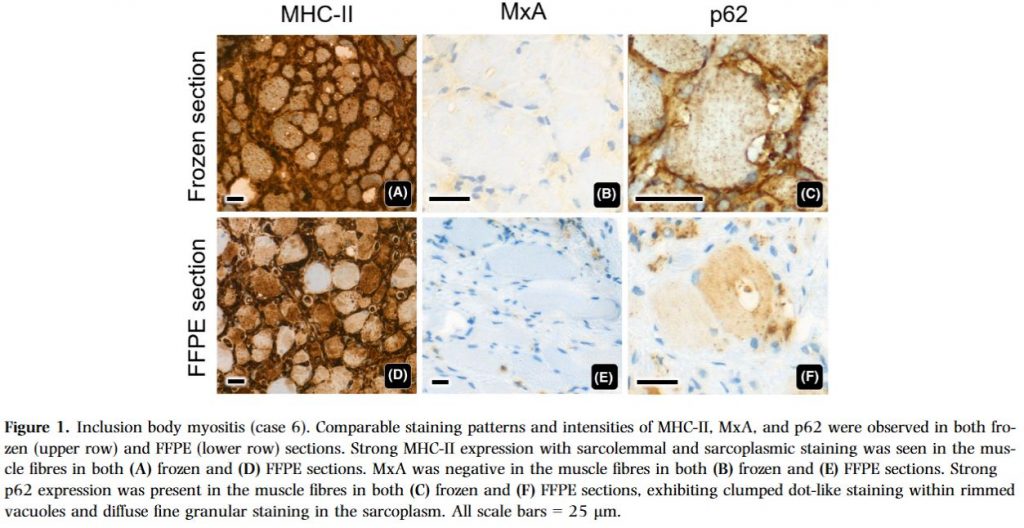
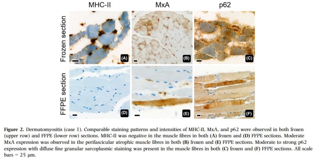
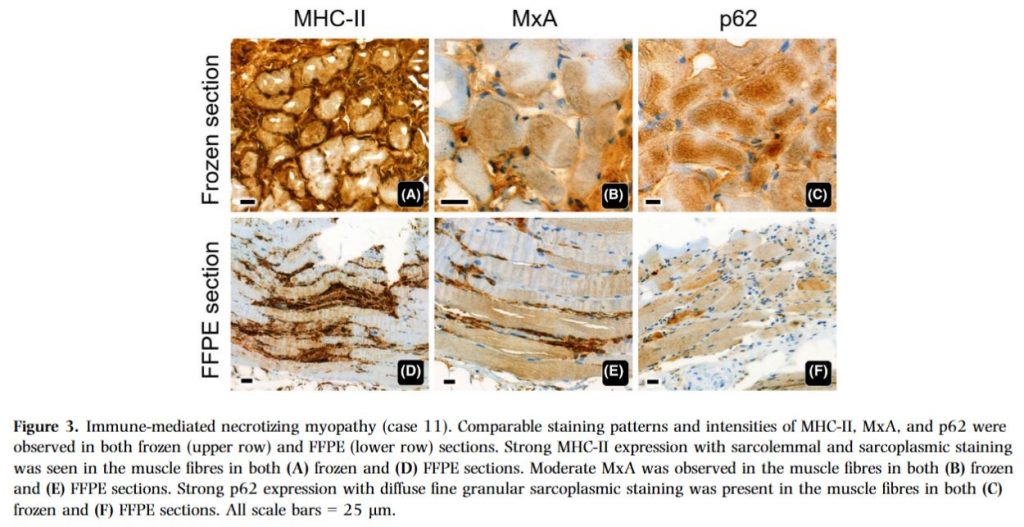
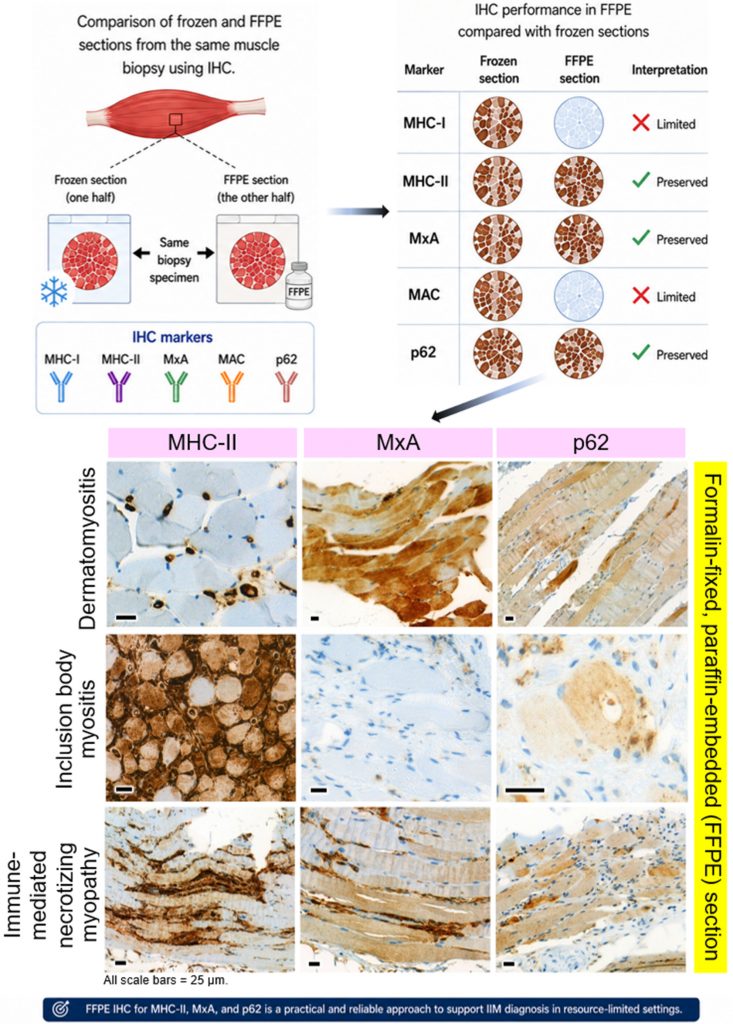
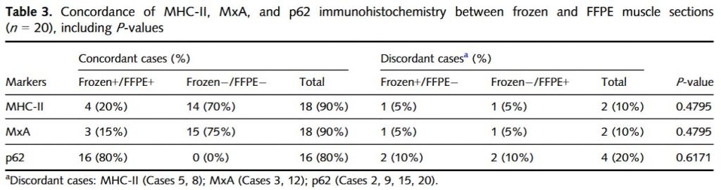
Comparable staining intensities and patterns for MHC-II, MxA, and p62 were achieved in FFPE muscle sections. Although discrepancies between frozen and FFPE sections occurred in eight cases, each involving a single marker, no statistically significant differences in immunohistochemical results were observed for MHC-II (P = 0.4795), MxA (P = 0.4795), and p62 (P = 0.6171). Concordance between methods was high, reaching 90% for MHC-II and MxA and 80% for p62. These isolated discrepancies may reflect variability in antigen expression associated with the segmental and patchy involvement characteristic of IIM. However, MHC-I and MAC staining were unsuccessful in FFPE sections.
Conclusions
Successful immunolabelling of MHC-II, MxA, and p62 was achieved in FFPE muscle sections. These findings indicate that several diagnostically informative markers for IIM can be reliably assessed in FFPE muscle, providing a practical diagnostic option in resource-constrained settings.
Keywords: Diagnostic immunohistochemistry, formalin-fixed and paraffin-embedded, idiopathic inflammatorymyopathy, muscle biopsy, snap-frozen section
Citation: Suriyonplengsaeng C, Waisayarat J. Diagnostic utility of inflammatory markers in formalin-fixed and paraffin-embedded muscle biopsies for idiopathic inflammatory myopathies. Histopathology. 2026 Jun 10. doi: 10.1111/his.70191. Epub ahead of print. PMID: 42266100.
RELATED SDGs:
15. LIFE ON LAND

ผู้ให้ข้อมูล: ผู้ช่วยศาสตราจารย์ นพ.ชินวุฒิ สุริยนเปล่งแสง
ชื่ออาจารย์ที่ทำวิจัย: ผู้ช่วยศาสตราจารย์ นพ.ชินวุฒิ สุริยนเปล่งแสง
แหล่งทุนวิจัย: This research was funded by the Research Grant for Retrospective Study (Grant Type 5), Faculty of Medicine Ramathibodi Hospital, Mahidol University, Bangkok, Thailand (Grant No. R1106905001).
เครดิตภาพ: ผู้ช่วยศาสตราจารย์ นพ.ชินวุฒิ สุริยนเปล่งแสง
Tags: Diagnostic immunohistochemistry, formalin-fixed and paraffin-embedded, idiopathic inflammatorymyopathy, muscle biopsy, snap-frozen section