Highlight
ความแตกต่างในการแสดงออกของ α-SMA และจำนวนเซลล์ที่แตกต่างกันของ medial collateral ligament ในส่วนต้นและส่วนปลายอาจอธิบายความสามารถในการรักษาที่แตกต่างกัน ซึ่งสนับสนุนความถูกต้องของทฤษฎี epiligament ที่ในการฟื้นตัวของเอ็นจากอาการบาดเจ็บ
ที่มาและความสำคัญ
หัวเข่าของมนุษย์เป็นข้อต่อที่ซับซ้อนที่ประกอบด้วยเอ็นหลายเส้น รวมถึง medial collateral ligament (MCL) ที่ให้ความมั่นคงแก่เข่าและช่วยป้องกันการเคลื่อนไหวเข้าด้านในมากเกินไป โดย MCL จะมีชั้นเนื้อเยื่อเกี่ยวพันที่เรียกว่า epiligament (EL) ซึ่งดึงดูดความสนใจในด้านการวิจัยในระยะหลังอย่างยิ่งเนื่องจากอาจมีการแสดงออกของโมเลกุลต่างๆที่ช่วยฟื้นฟูอาการบาดเจ็บของ MCL การศึกษาครั้งนี้มีวัตถุประสงค์เพื่อเปรียบเทียบการแสดงออกของ vascular endothelial growth factor (VEGF), CD34, and α-smooth muscle actin (α-SMA) ในส่วนต้นและส่วนปลาย MCL เพื่อทำความเข้าใจให้ดีขึ้นว่าโมเลกุลเหล่านี้ส่งผลต่อการรักษาการบาดเจ็บของเอ็นได้อย่างไร
Abstract
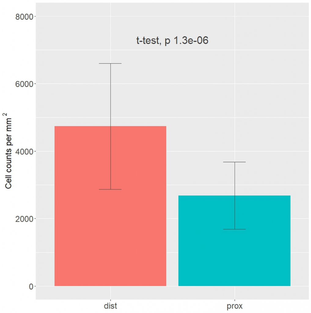
The human knee is a complex joint that comprises several ligaments, including the medial collateral ligament (MCL). The MCL provides stability to the knee and helps prevent its excessive inward movement. The MCL also has a thin layer of connective tissue known as the epiligament (EL), which adheres to the ligament. This unique feature has drawn attention in the field of ligament healing research, as it may have implications for the recovery process of MCL injuries. According to the EL theory, ligament regeneration relies heavily on the provision of cells, blood vessels, and molecules. The present study sought to compare the expression of vascular endothelial growth factor (VEGF), CD34, and α-smooth muscle actin (α-SMA) in healthy knees’ proximal and distal MCL segments to better understand how these proteins affect ligament healing. By improving the EL theory, the current results could lead to more effective treatments for ligament injury. To conduct the present analysis, monoclonal antibodies were used against CD34, α-SMA, and VEGF to examine samples from 12 fresh knee joints’ midsubstance MCLs. We identified a higher cell density in the EL than in the ligament connective tissue, with higher cell counts in the distal than in the proximal EL part. CD34 immunostaining was weak or absent in blood vessels and the EL, while α-SMA immunostaining was strongest in smooth muscle cells and the EL superficial layer. VEGF expression was mainly in the blood vessels’ tunica media. The distal part showed more SMA-positive microscopy fields and higher cell density than the proximal part (4735 vs. 2680 cells/mm2). Our study identified CD34, α-SMA, and VEGF expression in the MCL EL, highlighting their critical role in ligament healing. Differences in α-SMA expression and cell numbers between the ligament’s proximal and distal parts may explain different healing capacities, supporting the validity of the EL theory in ligament recovery.
KEYWORDS: medial collateral ligament, epiligament, theory, knee joint
Citation: Georgiev, G.P.; Yordanov, Y.; Gaydarski, L.; Tubbs, R.S.; Olewnik, Ł.; Zielinska, N.; Piagkou, M.; Ananiev, J.; Dimitrova, I.N.; Slavchev, S.A.; et al. Are There Any Differences in the Healing Capacity between the Medial Collateral Ligament’s (MCL) Proximal and Distal Parts in the Human Knee? Quantitative and Immunohistochemical Analysis of CD34, α-Smooth Muscle Actin (α-SMA), and Vascular Endothelial Growth Factor (VEGF) Expression Regarding the Epiligament (EL) Theory. Biomedicines2024, 12, 659.
DOI: https://doi.org/10.3390/biomedicines12030659
RELATED SDGs:
3. GOOD HEALTH AND WELL-BEING

ผู้ให้ข้อมูล: ผู้ช่วยศาสตราจารย์ ดร.อธิคุณ สุวรรณขันธ์
ชื่ออาจารย์ที่ทำวิจัย: ผู้ช่วยศาสตราจารย์ ดร.อธิคุณ สุวรรณขันธ์
Credit ภาพ: ผู้ช่วยศาสตราจารย์ ดร.อธิคุณ สุวรรณขันธ์
Webmaster: ว่าที่ ร.อ. นเรศ จันทรังสิกุล
Tags: epiligament, knee joint, medial collateral ligament, theory