Highlight
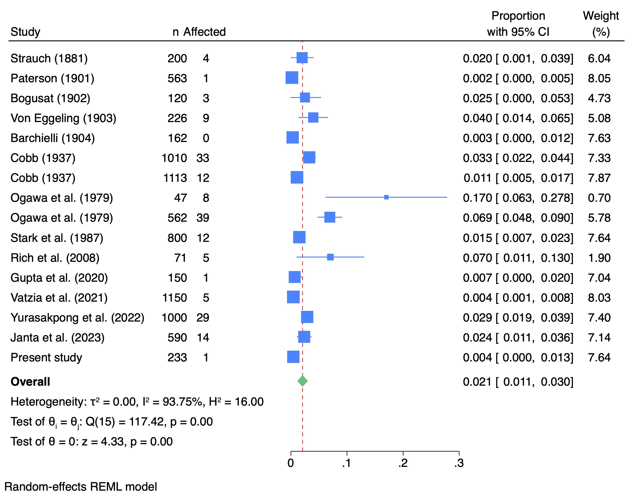
การศึกษานี้ประกอบด้วยการศึกษาจากภาพเอกซเรย์คอมพิวเตอร์ (computed tomography) และการวิเคราะห์อภิมานจากฐานข้อมูลอิเล็กทรอนิก 3 แหล่งได้แก่ Google Scholar, PubMed, and Journal Storage ผลงานวิจัยพบว่า จากคนไข้จำนวน 7,997 รายจากผลงานตีพิมพ์กว่า 16 ฉบับ episternal ossicles มีความชุกอยู่ที่ 2.1% โดยการศึกษาด้วย X-ray มีความชุกสูงสุดอยู่ที่ 7% และประชากรเอเชียมีความชุกของ episternal ossicles มากที่สุดที่ 3.8%


ที่มาและความสำคัญ
การมีอยู่ของ episternal ossicles ซึ่งเป็นโครงสร้างแปรผันทางกายวิภาคมีความสำคัญทางคลินิกเป็นอย่างยิ่งโดยเฉพาะกับรังสีแพทย์เพื่อป้องกันการวินิจฉัยผิดพลาด การศึกษาและการวิเคราะห์อภิมานฉบับนี้จัดทำขึ้นเพื่อศึกษาความชุกของ episternal ossicles ในประชากรสากล และศึกษาปัจจัยต่าง ๆ ที่ส่งผลต่อการมีอยู่ของโครงสร้างนี้
Abstract
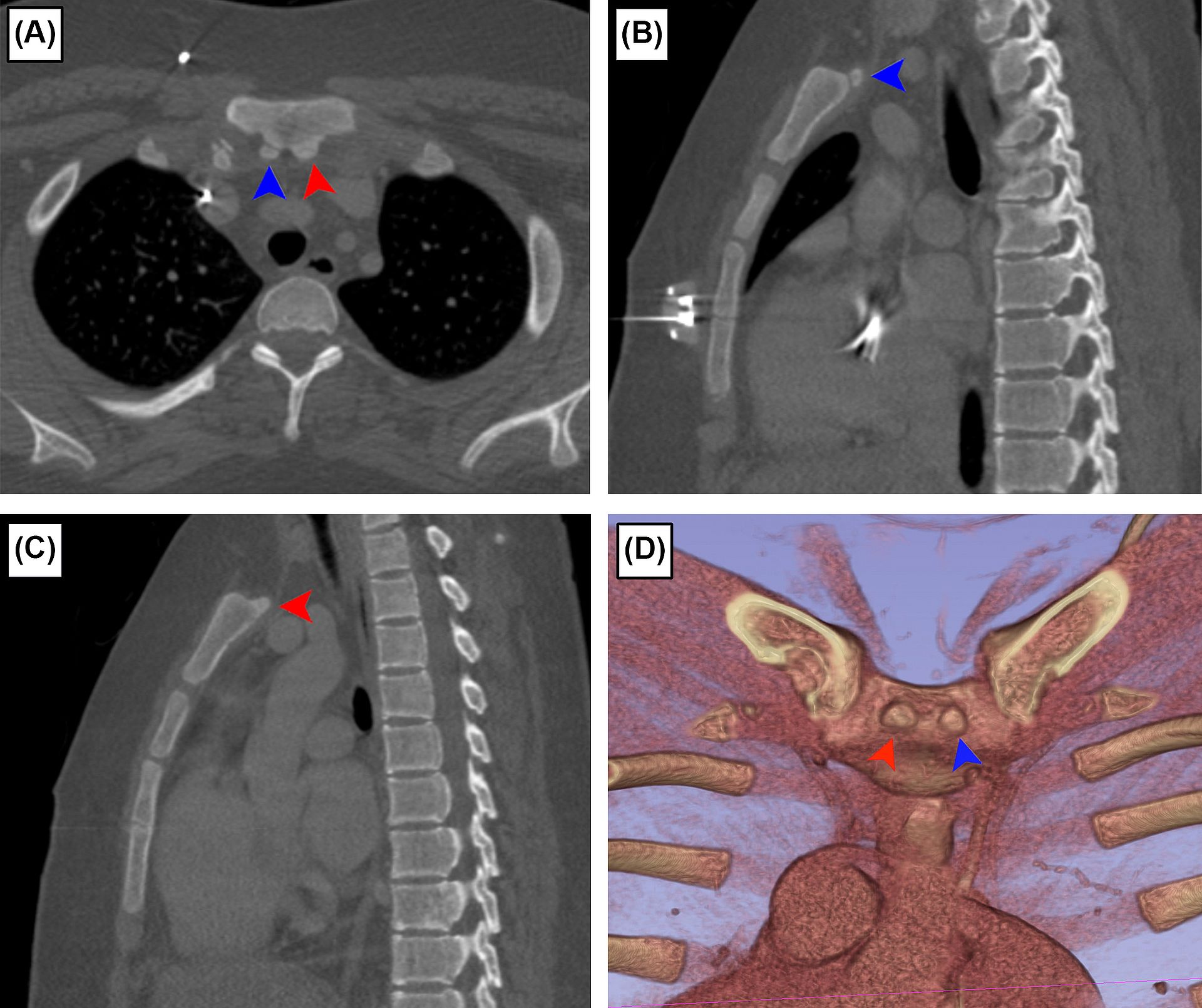
Episternal ossicles (EO) are accessory bones located superior and posterior to the manubrium, representing an anatomical variation in the thoracic region. This study aimed to investigate the prevalence and developmental aspects of EO in global populations. The prevalence of EO in pediatric populations was assessed using the “Pediatric-CT-SEG” open-access data set obtained from The Cancer Imaging Archive, revealing a single incidence of EO among 233 subjects, occurring in a 14-year-old patient. A meta-analysis was conducted using data from 16 studies (from 14 publications) through three electronic databases (Google Scholar, PubMed, and Journal Storage) encompassing 7997 subjects. An overall EO prevalence was 2.1% (95% CI 1.1–3.0%, I2 = 93.75%). Subgroup analyses by continent and diagnostic methods were carried out. Asia exhibited the highest prevalence of EO at 3.8% (95% CI 0.3–7.5%, I2 = 96.83%), and X-ray yielded the highest prevalence of 0.7% (95% CI 0.5–8.9%, I2 = 0.00%) compared with other modalities. The small-study effect was indicated by asymmetric funnel plots (Egger’s z = 4.78, p < 0.01; Begg’s z = 2.30, p = 0.02). Understanding the prevalence and developmental aspects of EO is crucial for clinical practitioners’ awareness of this anatomical variation.
KEYWORDS: Episternal ossicles, Sternum, Anatomical variation, Computed tomography, Systematic review, Meta-analysis
Citation: Pongruengkiat, W., Pitaksinagorn, W., Yurasakpong, L., Taradolpisut, N., Kruepunga, N., Chaiyamoon, A., & Suwannakhan, A. (2024). Anatomical study and meta-analysis of the episternal ossicles. Surgical and Radiologic Anatomy, 46, 195-202.
DOI: https://doi.org/10.1007/s00276-023-03280-y
RELATED SDGs:
3. GOOD HEALTH AND WELL-BEING

ผู้ให้ข้อมูล: ผู้ช่วยศาสตราจารย์ ดร.อธิคุณ สุวรรณขันธ์
ชื่ออาจารย์ที่ทำวิจัย: ผู้ช่วยศาสตราจารย์ ดร.อธิคุณ สุวรรณขันธ์, อาจารย์ ดร.ลภัสรดา ยุรศักดิ์พงศ์
ชื่อนักศึกษาที่ทำวิจัย: นางสาวนภาวรรณ ธาราดลพิสุทธิ์
Credit ภาพ: ผู้ช่วยศาสตราจารย์ ดร.อธิคุณ สุวรรณขันธ์
Webmaster: ว่าที่ ร.อ. นเรศ จันทรังสิกุล
Tags: anatomical variation, computed tomography, Episternal ossicles, meta-analysis, Sternum, systematic review
