Highlight:
งานวิจัยนี้ใช้เทคโนโลยีสามมิติเพื่อรายงานโครงสร้างผันแปรโครงสร้างหนึ่ง (a reverse form of Linburg–Comstock variation) ซึ่งผู้อ่านสามารถมีปฏิสัมพันธ์กับโครงสร้างนี้ได้โดยตรง การรายงานผลงานวิจัยแบบใหม่นี้อาจเป็นบรรทัดฐานใหม่ของการนำเสนอข้อมูลทางกายวิภาคในอนาคต
ที่มาและความสำคัญ
ปัจจุบันการสแกนภาพแบบสามมิติ เป็นเทคโนโลยีที่มีการนำไปใช้อย่างแพร่หลาย โดยเฉพาะในด้านการศึกษา ที่ผ่านมาได้มีการนำเทคโนโลยีการสแกนภาพสามมิติมาใช้จัดทำเป็นสื่อประกอบการเรียนการสอนกายวิภาคศาสตร์ และพบว่าส่งผลให้ผู้เรียนสามารถจดจำเนื้อหาและระบุตำแหน่งของโครงสร้างต่าง ๆ ได้ดียิ่งขึ้น ซึ่งนอกจากเป็นการเพิ่มประสิทธิภาพให้กับการเรียนการสอนแล้วยังเป็นประโยชน์อย่างยิ่งสำหรับสถาบันการศึกษาที่กำลังประสบปัญหาขาดแคลนร่างอาจารย์ใหญ่หรือไม่สามารถดำเนินการจัดการเรียนการสอนแบบ on-site ได้ในช่วงสภาวะการระบาดของโรคโควิด-19 นี้ อย่างไรก็ตาม การนำเทคโนโลยีสแกนภาพสามมิติมาใช้ในเชิงวิจัยยังมีไม่มากนัก โดยเฉพาะอย่างยิ่งการนำภาพสแกนสามมมิติมาใช้เพื่อเป็นเครื่องมือในการนำเสนอผลงานวิจัยทางกายวิภาคศาสตร์ ยังไม่เคยพบว่ามีการนำมาใช้ ซึ่งปกติงานวิจัยทางด้านกายวิภาคศาสตร์จะใช้การนำเสนอ รายงานผล แบบรูปภาพสองมิติ (2D) หรือมีการวาดภาพประกอบร่วมด้วย ดังนั้น ในงานวิจัยฉบับนี้ ผู้วิจัยได้ทดลองการนำเสนอรายงานผลการวิจัย โดยใช้เครื่องสแกนภาพแบบสามมิติ (3D scanner) เพื่อสแกนแขนท่อนล่างของร่างอาจารย์ใหญ่เพื่อนำมารายงานโครงสร้างแปรผันทางกายวิภาค ซึ่งเป็นเส้นเอ็นที่เชื่อมระหว่างกล้ามเนื้อ flexor pollicis longus และกล้ามเนื้อ flexor digitorum profundus ของนิ้วชี้ (a reverse form of Linburg–Comstock variation) และนำไปสร้างเป็นไฟล์ 3D-PDF โดยผู้อ่านสามารถหมุน ขยาย และมีปฏิสัมพันธ์กับโครงสร้างนั้น ๆ ได้ ในลักษณะเดียวกับการปฏิบัติในห้องปฏิบัติการ ด้วยจุดเด่นเหล่านี้ผู้วิจัยเล็งเห็นว่าการรายงานผลงานวิจัยในรูปแบบสามมิติอาจกลายเป็นบรรทัดฐานใหม่ของการนำเสนอข้อมูลทางกายวิภาคศาสตร์ในอนาคตอันใกล้นี้ นอกจากนี้ ผู้วิจัยยังได้เสนอสมมติฐานซึ่งอธิบายกลไกลการเกิดขึ้นของโครงสร้างนี้ในเชิงวิวัฒนาการและวิเคราะห์ความสำคัญทางคลินิกของโครงสร้างนี้อีกด้วย
Abstract
Purpose
Two most common variations of flexor pollicis longus include its accessory head and its connection with the flexor digitorum profundus of the index (Linburg–Comstock variation). In addition, while three-dimensional (3D) screening has widely been used in anatomical education, its use as reporting tool in anatomical research is still limited. The objective of this study is to report a previously unrecognized form of the accessory head of flexor pollicis longus, discuss the potential etiology of Linburg–Comstock variation, and pilot the 3D scanning of a large-scale anatomical structure.
Methods
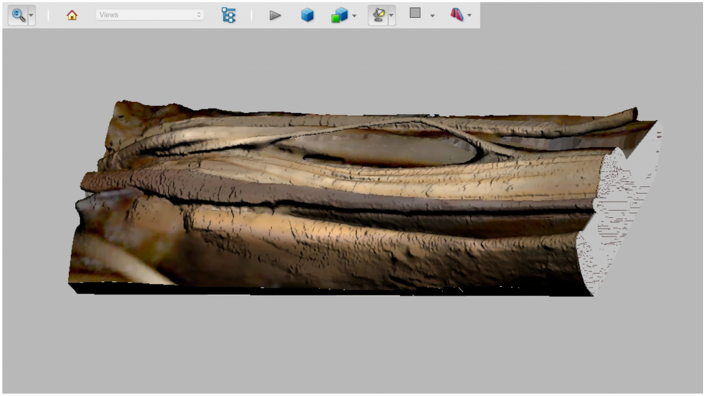
An unusual tendon slip was discovered during a routine dissection in the anterior compartment of the right forearm of a 54-year-old male cadaver. A 3D scanner was used to capture the surface topography of the specimen and an interactive portable document format (PDF) was created.
Results
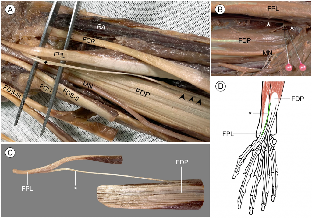
An anomalous tendon was found originating from the lateral aspect of the flexor digitorum profundus muscle. This variant tendon then inserted onto the medial surface of the flexor pollicis longus tendon before entering the carpal tunnel. The variation resembles a reverse form of Linburg–Comstock variation, because pulling this variant tendon resulted in simultaneous flexion of the interphalangeal joint of thumb.
Conclusion
Surgeons should be aware of the reverse Linburg–Comstock variation, because it may not be detectable by the conventional provocative testing. Linburg–Comstock variation may be classified as an anatomical variant or a secondarily acquired condition depending on its type. Our demonstration of interactive 3D-PDF file highlights its potential use for delivering anatomical information in future cadaveric studies.
KEYWORDS: Flexor pollicis longus, Flexor digitorum profundus, Linburg–Comstock variation, Anatomical variations, 3D scanning, 3D visualization, Surface rendering
Citation: Prasatkaew, W., Kruepunga, N., Yurasakpong, L. et al. A reverse form of Linburg–Comstock variation with comments on its etiology and demonstration of interactive 3D portable document format. Surg Radiol Anat (2021). https://doi.org/10.1007/s00276-021-02858-8
RELATED SDGs:
SDG Goal หลัก ที่เกี่ยวข้อง
4. QUALITY EDUCATION

SDG Goal ที่เกี่ยวข้องอื่น ๆ
3. GOOD HEALTH AND WELL-BEING

ผู้ให้ข้อมูล: นางวิจิตรา ประสาทแก้ว
ชื่อบุคลากรและอาจารย์ที่ทำวิจัย: นางวิจิตรา ประสาทแก้ว อาจารย์ ดร.อธิคุณ สุวรรณขันธ์ อาจารย์ ดร.ณัฐเมธี เครือภูงา
ชื่อนักศึกษาที่ทำวิจัย: น.ส.ลภัสรดา ยุรศักดิ์พงศ์
ภาพถ่าย: อาจารย์ ดร.ณัฐเมธี เครือภูงา, อาจารย์ ดร.อธิคุณ สุวรรณขันธ์, นางวิจิตรา ประสาทแก้ว