Highlight
งานวิจัยฉบับนี้ศึกษาความชุกและสัณฐานของรูเปิดบนกระดูกสะบักหรือ scapular foramina และใช้การสแกนสามมิติและเทคโนโลยี AR ซึ่งเป็นมิติใหม่ของการนำเสนอผลงานวิจัยทางกายวิภาคและอาจเป็นมาตรฐานใหม่ในอนาคต
ที่มาและความสำคัญ
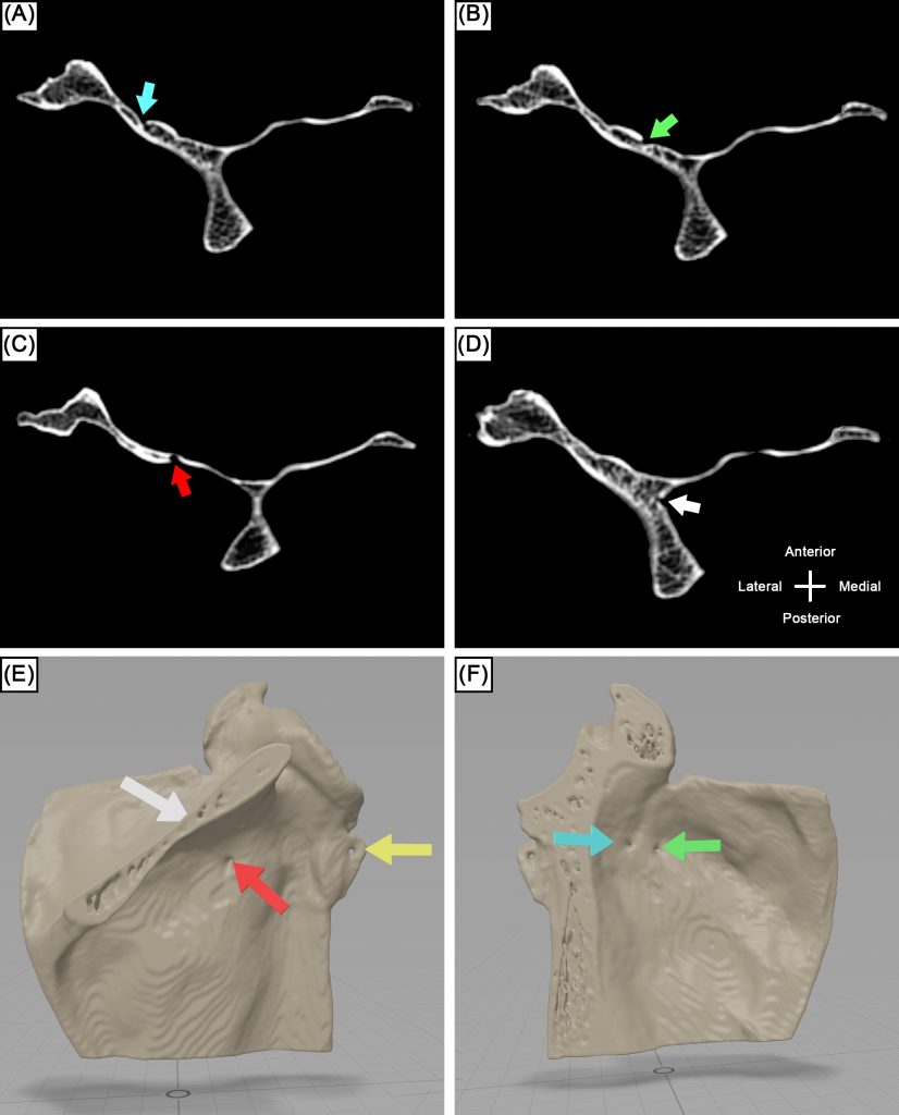
การมีอยู่ของรู scapular foramina อาจสับสนกับการหักของกระดูกสะบักเมื่อวินิจฉัยจากภาพรังสี การรับรู้ถึงตำแหน่งของ scapular nutrient foramina มีความสำคัญเป็นอย่างยิ่งเพื่อป้องกันความเสียหายต่อหลอดเลือด nutrient vessels เมื่อทำหัตการบริเวณกระดูกสะบัก งานวิจัยฉบับนี้ศึกษาความชุกและสัณฐานของรูเปิดและรูสารอาหารของกระดูกสะบัก โดยใช้กระดูกสะบักจำนวนทั้งสิ้น 150 ชิ้น การศึกษาก่อนหน้าพบว่ารูเปิดบนกระดูกสะบัก หรือ scapular foramina เป็นรูเปิดที่เชื่อมพื้นผิวแต่ละด้านของกระดูกสะบัก ซึ่งเป็นโครงสร้างแปรผันทางกายวิภาค (anatomical variant) ที่มีความชุกเพียงร้อยละ 7.5 ของประชากร อย่างไรก็ตามงานวิจัยฉบับนี้พบว่า scapular foramina นั้นมีความชุกสูงถึงร้อยละ 78 ของประชากรเมื่อศึกษาด้วยเส้นลวดขนาดเล็ก (0.36 มม.) มีรูปแบบของการเชื่อมต่อถึงกว่า 18 แบบและอาจมีรูเปิดได้มากถึง 4 รู ซึ่งมองเห็นได้ชัดเจนจากภาพเอกซเรย์ computed tomography ด้วยความชุกกว่าร้อยละ 78 จึงสรุปได้ว่า scapular foramina นั้นแท้จริงแล้วเป็นโครงสร้างปกติแทนที่จะเป็นโครงสร้างแปรผัน
Abstract
Purpose
The aim of our study is to study the prevalence and anatomy of scapular foramina (SF) and scapular nutrient foramina (SNF) in dried skeletons from the Northeastern Thai population.
Methods
A total of 150 dried scapulae were investigated. Both SF and SNF were identified using a metal wire with a diameter of 0.36 mm. The number, locations, lengths, and diameters of SF were recorded. Subsequently, SNF were identified using the same metal wire. Their number and locations were recorded. Two observers performed the evaluations and measurements.
Results
SF were present in 78.0% of scapulae. They could have up to five openings. Eighteen types were found. On average they were longer in males (21.7 ± 5.0 mm) than females (19.45 ± 4.6 mm). The mean diameters of both the superior and inferior openings were significantly greater in females (p < 0.01). SNF, in contrast, were present in 100% of scapulae. They were located in the supraspinous fossa (36.7%), subscapular fossa (31.3%), infraspinous fossa (22.8%), and peri-glenoid area (10.0%).
Conclusion
Unlike previous studies, the present study suggests that SF are normal anatomical findings, present in 78.0% of the scapulae investigated. Surgeons should be aware of both SNF and SF when operating or interpreting radiological findings.
KEYWORDS: Scapula, Scapular nutrient foramen, Scapular foramen, Anatomical variations
Citation: Yurasakpong, L., Suwannakhan, A., Kirisattayakul, W., Samrid, R., Iamsaard, S., Limwachiranon, J., Khanthiyong, B., Tubbs, R., Iwanaga, J. & Chaiyamoon, A. (2023). Topographical study of scapular foramina and scapular nutrient foramina in dried skeletons. Surgical and Radiologic Anatomy, 45(5), 563-570. https://doi.org/10.1007/s00276-023-03132-9
RELATED SDGs:
3. GOOD HEALTH AND WELL-BEING

ผู้ให้ข้อมูล: ผู้ช่วยศาสตราจารย์ ดร.อธิคุณ สุวรรณขันธ์
ชื่ออาจารย์ที่ทำวิจัย: อาจารย์ ดร.ลภัสรดา ยุรศักดิ์พงศ์, ผู้ช่วยศาสตราจารย์ ดร.อธิคุณ สุวรรณขันธ์
Credit ภาพ: ผู้ช่วยศาสตราจารย์ ดร.อธิคุณ สุวรรณขันธ์
Tags: anatomical variation, Mandible, meta-analysis, Stafne bone cavity, systematic review