Highlight
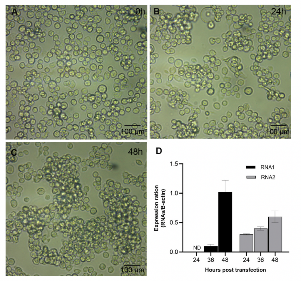
นักวิทยาศาสตร์สามารถจำลองการแบ่งตัวและการเพิ่มจำนวนของไวรัสได้ในระบบเพาะเลี้ยงกับเซลล์แมลง ซึ่งมีข้อดีในการเพิ่มจำนวนไวรัสได้จำนวนมาก ผู้วิจัยจึงได้นำระบบเพาะเลี้ยงดังกล่าวมาใช้ในการผลิตและเพิ่มจำนวนไวรัส MrNV ในเซลล์แมลง S2 โดยการจำลองการติดเชื้อเข้าไปในเซลล์ S2 เป็นเวลา 24 ชั่วโมง เพื่อเก็บน้ำเพาะเลี้ยงเซลล์ที่มีอนุภาคไวรัส MrNV ที่ถูกปลดปล่อยออกมา แล้วนำเชื้อดังกล่าวมาทดสอบการติดเชื้อในเซลล์ S2 ใหม่อีกรอบ เพื่อทดสอบความสามารถของไวรัสในการติดเชื้อซ้ำ พบว่ามีการปรากฏของไวรัสใหม่ในเซลล์ S2 และก่อให้เกิดการเปลี่ยนแปลงของเซลล์เช่นเดียวกับไวรัสในธรรมชาติ นอกจากนี้ หากมีการฉีดเชื้อเข้าไปในกุ้งแชบ๊วย (P. merguiensis) จะก่อให้เกิดอาการกล้ามเนื้อขาวภายใน 24 ชั่วโมง และเกิดการลดลงของจำนวนเม็ดเลือดในช่วง 6-24 ชั่วโมงก่อนที่กลับมาเป็นปกติภายใน 48 ชั่วโมง รวมถึงการเพิ่มจำนวนยีนที่เกี่ยวข้องกับภูมิคุ้มกันกุ้ง เช่น ยีน HSP70 และยีน trypsin เป็นต้น จึงสรุปได้ว่า เราสามารถสร้างไวรัส MrNV ในระบบเพาะเลี้ยงเซลล์แมลง S2 เพื่อนำมาใช้ประโยชน์ต่อไปในการศึกษากลไกการติดเชื้อ และพัฒนาต่อยอดในการหายุทธวิธีในการยับยั้ง/รักษาโรคติดเชื้อไวรัสในกุ้งต่อไป

ชื่องานวิจัยภาษาไทย
การทดสอบการติดเชื้อและความรุนแรงของไวรัสโนดาจากกุ้งก้ามกรามที่ผลิตจากเซลล์แมลงหวี่เมลาโนกาลเตอร์ และการทดสอบการติดเชื้อในกุ้งแชบ๊วย
Abstract
It has been established that baculovirus-insect cell line is applicable for shrimp virus replication, propagation and secretion in the in vitro culture system. We thus aimed to produce Macrobrachium rosenbergii nodavirus (MrNV) clone within S2 cell to improve viral production over the previous model using Sf9 cell. Upon the transfection of genomic RNA1 and RNA2 into S2 cells, the recognizable cellular changes including cytoplasmic swelling and clumping of cells were observed within 24 h. The culture media containing secreted MrNV particles were re-transfected into healthy S2 cells and similar cellular changes as with the first transfection were observed. Immunohistochemistry analysis of the re-infecting S2 cell revealed an intense immunoreactivity against MrNV capsid protein confirming that S2 cell was permissive cells for MrNV. In vivo infectivity test using P. merguiensis as a model animal exposed to the secreted MrNV revealed the presence of RNA2 fragment in shrimp tissue accompanied with the sign of whitish abdominal muscle at 24 h post-infection (p.i.). In addition, the number of shrimp hemocytes decreased at 6–24 h p.i. and returned to the normal level at 48 h p.i., whereas a significant up-regulation of immune-related genes including HSP70 and trypsin was noted. These data suggested that rescued MrNV produced in S2 is practically useful for MrNV infection test in which their natural virion inoculae are difficult to obtain. In addition, the molecular basis of viral pathogenesis can further be investigated which should be beneficial for any antiviral therapy developments in the future.
ที่มาและความสำคัญ
การหาระบบการเพาะเลี้ยงไวรัสให้ได้ปริมาณมากๆจากเซลล์แมลงหวี่ โดยที่ไวรัสยังมีคุณสมบัติและความรุนแรงในการก่อโรคเท่ากับไวรัสธรรมชาติ
KEYWORDS: Drosophila melanogaster, Immune system, Macrobrachium rosenbergii nodavirus, Penaeus merguiensis
Citation:
Weerachatyanukul W, Pooljun C, Hirono I, Chotwiwatthanakun C, Jariyapong P. Infectivity and virulence of the infectious Macrobrachium rosenbergii nodavirus produced from Drosophila melanogaster cell using Penaeus merguiensis as an infection model. Fish Shellfish Immunol. 2023 Jan;132:108474. doi: 10.1016/j.fsi.2022.108474. Epub 2022 Dec 5. PMID: 36481289.
RELATED SDGs:
SDG Goal หลัก ที่เกี่ยวข้อง
2. ZERO HUNGER

SDG Goal ที่เกี่ยวข้องอื่น ๆ
8. DECENT WORK AND ECONOMIC GROWTH

ผู้ให้ข้อมูล: รองศาสตราจารย์ ดร.วัฒนา วีรชาติยานุกูล
ชื่ออาจารย์ที่ทำวิจัย: รองศาสตราจารย์ ดร.วัฒนา วีรชาติยานุกูล
แหล่งทุนวิจัย: National Research Council of Thailand (Grant No. NRCT5-RSA63019-05 to PJ and NRCT5-RSA63015-02 to WW)
ภาพถ่าย: รองศาสตราจารย์ ดร.วัฒนา วีรชาติยานุกูล
Tags: Drosophila melanogaster, Immune system, Macrobrachium rosenbergii nodavirus, Penaeus merguiensis
