Highlight
การศึกษานี้แสดงให้เห็นว่าแบบจำลองหนูพาร์กินสันที่เหนี่ยวนำด้วย MPTP มีการเสื่อมของเซลล์ประสาทโดปามิเนอร์จิก ร่วมกับความผิดปกติของการทรงตัวและการเคลื่อนไหว นอกจากนี้ยังพบการเปลี่ยนแปลงของฟอสโฟลิพิด โดยเฉพาะการลดลงของ phosphatidylcholine ที่มีกรดไขมันไม่อิ่มตัวเชิงซ้อนในสมอง ซึ่งสัมพันธ์กับการสูญเสียเซลล์ประสาทและอาการทางพฤติกรรม สะท้อนบทบาทสำคัญของไขมันในพยาธิกำเนิดของโรคพาร์กินสัน
ที่มาและความสำคัญ
โรคพาร์กินสันเป็นโรคทางระบบประสาทเสื่อมที่พบมากในผู้สูงอายุ ส่งผลให้เกิดความผิดปกติของการเคลื่อนไหว โดยมีสาเหตุหลักจากการเสื่อมของเซลล์ประสาทโดปามิเนอร์จิกในสมอง แม้ว่าพยาธิสภาพของโรคจะเกี่ยวข้องกับการสะสมของโปรตีนผิดปกติ เช่น Lewy bodies แต่ปัจจุบันพบว่าเมแทบอลิซึมของไขมัน โดยเฉพาะกรดไขมันไม่อิ่มตัวเชิงซ้อนก็มีบทบาทสำคัญต่อการเกิดโรค
งานวิจัยที่ผ่านมาแสดงให้เห็นการเปลี่ยนแปลงของไขมันในสมองผู้ป่วยพาร์กินสัน อย่างไรก็ตาม เทคนิคที่ใช้ยังไม่สามารถแสดงการกระจายตัวของไขมันในเนื้อเยื่อได้อย่างชัดเจน ดังนั้น การใช้เทคนิค MALDI-MSI ซึ่งสามารถวิเคราะห์ตำแหน่งของไขมันในเนื้อเยื่อโดยตรง จึงมีความสำคัญในการศึกษาการเปลี่ยนแปลงของไขมันในบริเวณ motor cortex ซึ่งเกี่ยวข้องกับการควบคุมการเคลื่อนไหว
การศึกษานี้จะช่วยเพิ่มความเข้าใจกลไกระดับโมเลกุลของโรคพาร์กินสัน และอาจนำไปสู่แนวทางใหม่ในการวินิจฉัยและรักษาในอนาคต
Abstract
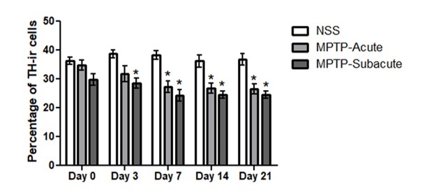
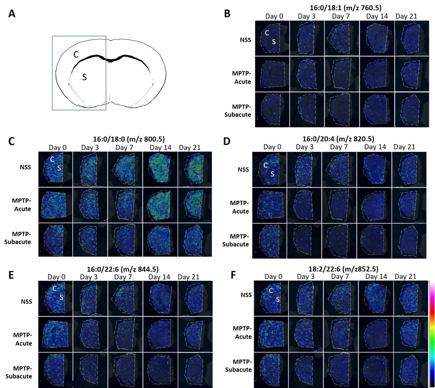
Parkinson’s disease (PD) is a neurodegenerative disorder caused by the death of dopaminergic neurons in the substantia nigra pars compacta (SNc). Lipid metabolism, especially phospholipids, has been reported to be altered in PD. The purpose of this study is to investigate the temporal expression and spatial distribution of phospholipids in the motor cortex and striatum at different time points of PD using matrix-assisted laser desorption/ionization mass spectrometry imaging (MALDI-MSI) in a 1-methyl-4-phenyl-1,2,3,6-tetrahydropyridine (MPTP)-induced parkinsonian mouse model. Mice were injected with saline (NSS) or MPTP at two different time points to create acute and subacute models. Motor analysis was performed at 0, 3, 7, 14, and 21 days post-injection. Tyrosine hydroxylase (TH) staining revealed progressive damage of neurons in the substantia nigra compacta (SNc) and reduced striatal fibers in MPTP-treated animals. By using MALDI-MSI, we identified changes in phosphatidylcholine (PC) profiles in the brains of MPTP-treated animals. Polyunsaturated PCs, including PC 36:4 (16:0/20:4), PC 38:6 (16:0/22:6), and PC 40:8 (18:2/22:6), were decreased in the MPTP-treated groups. These reductions were time-dependent and were more pronounced in the subacute MPTP-treated group. The loss of dopamine neurons caused by MPTP may be associated with the selective loss of polyunsaturated PCs in brain membranes, indicating that lipid metabolism and membrane structural alterations may contribute to the pathology of PD.
KEYWORDS: Parkinson’s disease, MPTP, lipid alterations, polyunsaturated fatty acids, MALDI-MSI
Citation: Sroyraya, M., Noonong, K., Sobhon, P., Siangcham, T., Waiyaput, W., Sansri, V., Chaithirayanon, K., & Chonpathompikunlert, P. (2026). Alterations in Phospholipid Levels and Spatial Distribution in the Motor Cortex and Their Correlation with Motor Performance in an MPTP-Induced Parkinsonian Mouse Model. Molecules, 31(7), 1175. https://doi.org/10.3390/molecules31071175
RELATED SDGs:
SDG Goal หลัก ที่เกี่ยวข้อง
3. GOOD HEALTH AND WELL-BEING

ผู้ให้ข้อมูล: ผู้ช่วยศาสตราจารย์ ดร.มรกต สร้อยระย้า
ชื่ออาจารย์ที่ทำวิจัย: ผู้ช่วยศาสตราจารย์ ดร.มรกต สร้อยระย้า รองศาสตราจารย์ ดร.กุลธิดา ชัยธีระยานนท์
แหล่งทุนวิจัย: Thailand Research Fund (TRF)
Credit ภาพ: ผู้ช่วยศาสตราจารย์ ดร.มรกต สร้อยระย้า
Webmaster: ว่าที่ ร.อ. นเรศ จันทรังสิกุล